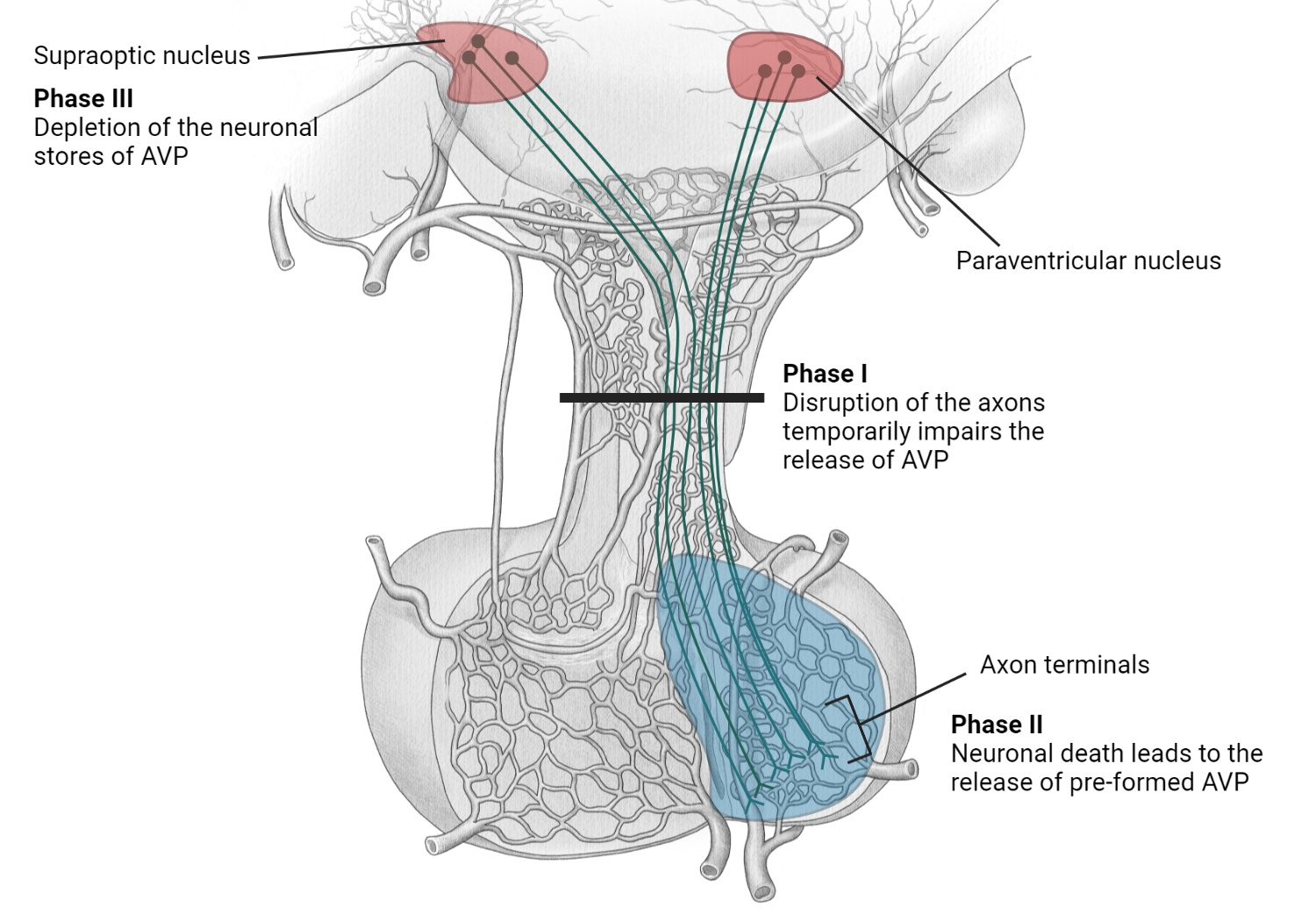
Complete transection of the pituitary stalk, as might occur in traumatic brain injury or transsphenoidal surgery, results in the classic “triphasic response” of central diabetes insipidus.
The triphasic response of central diabetes insipidus
- There is an initial phase of transient central diabetes insipidus, which occurs due to axonal shock and the inability of action potentials to be transferred from the cell body to the axon terminals in the neurohypophysis.
- The first phase is followed within 24 to 48 hours by the second phase of Syndrome of Inappropriate Antidiuretic Hormone (SIADH), due to axonal injury and rapid release of preformed arginine vasopressin (AVP) into circulation. This phase is sometimes referred to as the “normal” interphase. However, it is anything but normal since the release of vasopressin is largely unregulated and dependent on storage levels in the neurohypophysis. Indeed, the posterior pituitary has enough AVP to maintain water conservation for up to a week (an evolutionary adaptation).
- The third phase of recurrence of central diabetes insipidus occurs after the complete depletion of neuronal reserves of AVP. This final course of diabetes insipidus may be permanent, partial, or clinically silent. The challenge in managing this condition is realizing the clinical timeline of the triphasic response and altering fluid management promptly to prevent acute sodium imbalance.
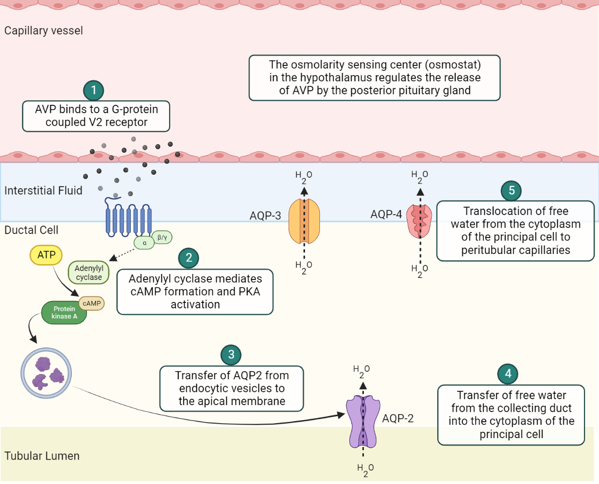
Role of AVP in water conservation
AVP binds to the G-protein-coupled V2 receptor on the basolateral membrane of the principal cell (step 1). This is followed by the activation of cytosolic adenylate cyclase, which subsequently increases cyclic AMP (cAMP) production (step 2). Increased intracellular cAMP activates Protein Kinase A (PKA) (step 3), which then facilitates the phosphorylation of AQP2 in its endocytic vesicles (step 4). AQP2 liberated from endocytic vesicles are translocated to the apical membrane (AM) of the principal cell (step 5). AQP3 and AQP4 are constitutively expressed (not released from endocytic vesicles) on the basolateral membrane, independent of PKA action. AQP2 moves free water from the collecting tubule into the cytosol of the ductal cell, while AQP3 and AQP4 water channels mediate the movement of free water across the basolateral membrane into the peritubular capillaries (step 6)
How does central diabetes insipidus lead to dehydration?
Down-regulation of vasopressin V2 receptors in the principal cell of the collecting tubule occurs due to the absence of trophic stimulation by Arginine Vasopressin (AVP), in the setting of complete central diabetes insipidus. Also, a loss of AVP stimulation causes internalization of aquaporin-2 (AQP2) water channels into cytosolic vesicles, which ultimately leads to free water loss
References
- Boone M, Deen PMT. Physiology and pathophysiology of the vasopressin-regulated renal water reabsorption. Pflugers Arch. 2008;456(6):1005-1024. doi:10.1007/s00424-008-0498-1
- Oksche A, Rosenthal W. The molecular basis of nephrogenic diabetes insipidus. J Mol Med Berl Ger. 1998;76(5):326-337.
Images(s) Courtesy
MyEndoConsult
Kindly Let Us Know If This Was helpful? Thank You!