Anatomy (Ultrasound views)
PART I : Anatomy of the neck and thyroid gland (ultrasound views)
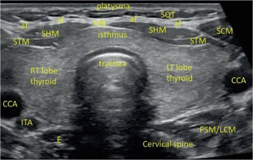
Fig 1.0 Transverse view of the thyroid gland showing relevant anatomic relations of the thyroid gland.
SQT = Subcutaneous tissue
AF = Anterior layer of the deep cervical fascia
SCM = Sternocleidomastoid muscle
SHM = Sternohyoid muscle
STM = Sternothyroid muscle
MR = Midline raphe for the fascia surrounding the SHM and STM muscles.
CCA = Common carotid artery
ITA = Inferior thyroid artery
IJV = Internal jugular vein
E = esophagus
PSM/LCM = Paraspinal muscle/longus colli muscle
Fig 2.0 Doppler ultrasound showing the left thyroid lobe and it’s relation to the internal jugular vein (IJV), Internal Carotid Artery (large pulsating vessel in the middle) and inferior thyroid artery.
The esophagus may lie behind either the left or right thyroid lobe. It can be errorneously identified as a thyroid nodule. Asking the patient to swallow will show areas of peristalsis.
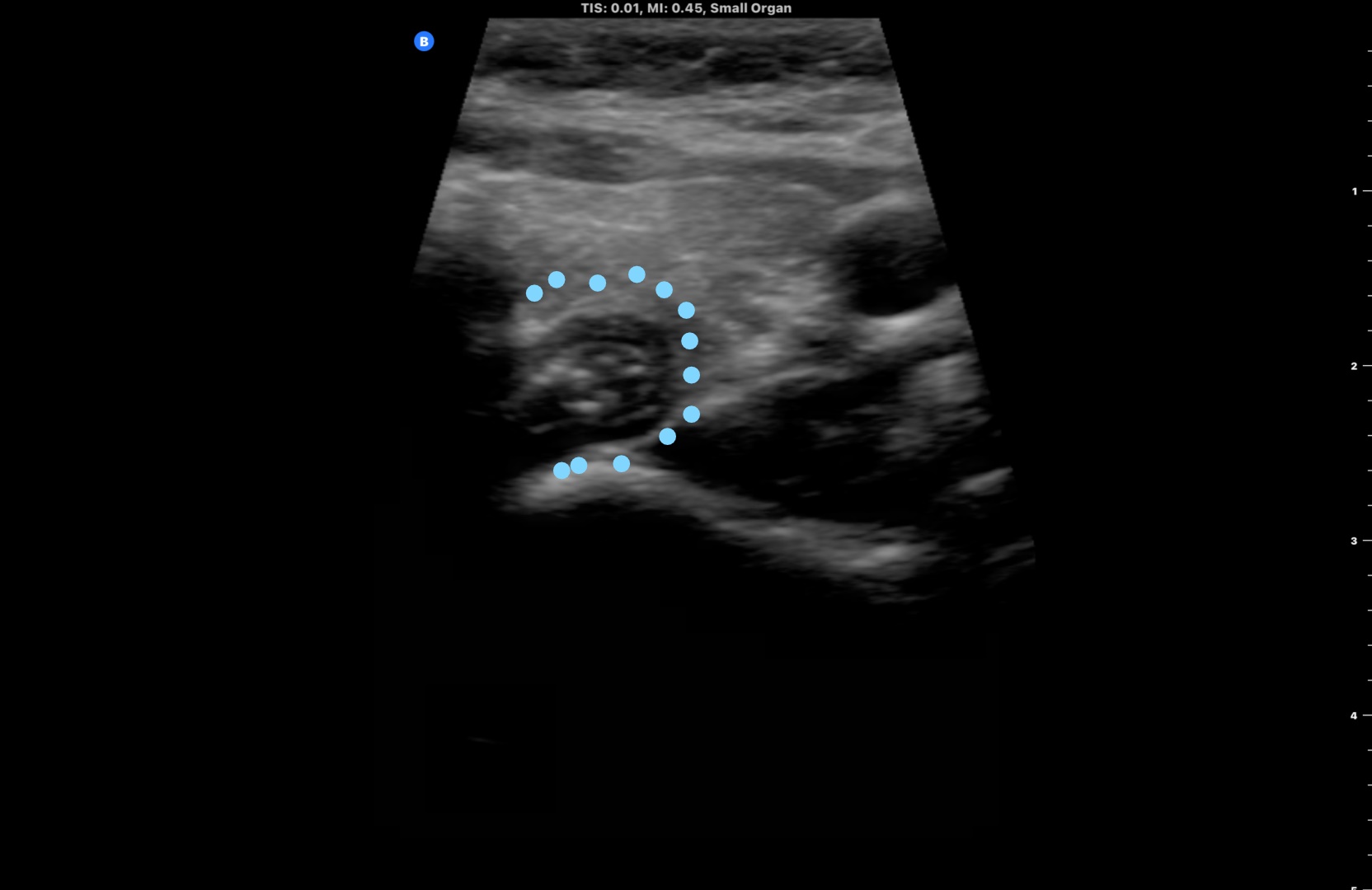
Fig 3.0 Identifying the esophagus (outlined by dots).
Figure 4.0 Peristalsis of the esophagus noted on deglutition.