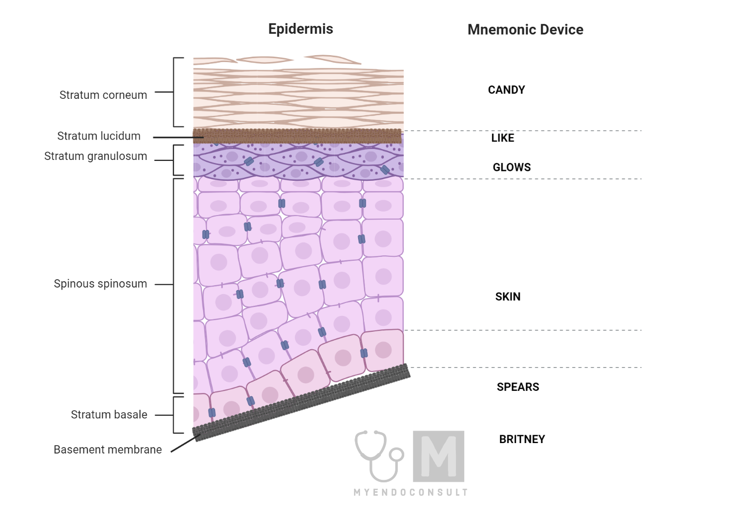
The complexity of the human body extends even to its exterior: the skin. This protective barrier is composed of various layers, each performing a distinct function. This article elucidates the five primary layers of the skin’s epidermis, from the base to the top: Stratum basale, Stratum spinosum, Stratum granulosum, Stratum lucidum, and Stratum corneum. We also provide mnemonic devices to aid memory, alongside detailed cellular and functional descriptions of each layer.
Introduction
The skin, beyond its protective role, has a highly organized structure with several layers, each exhibiting unique cellular characteristics and functions. Understanding these layers is critical in areas like dermatology, wound healing, and skin cancer research. Mnemonics are often used to aid memory, and we propose the mnemonic “Britney Spears Skin Glows Like Candy” from bottom to top.
Layers of the Skin
- Stratum Basale (Basal Layer): The Stratum basale, composed of columnar epithelial cells, is the wellspring of the skin’s layers. This basal layer houses cells that multiply and give rise to keratinocytes, which ascend through the layers. Melanocytes also reside here, producing and
- Stratum Spinosum (Prickle Cell Layer): The Stratum spinosum is characterized by polygonal cells connected via desmosomal bridges, lending this layer its ‘prickly’ appearance.
- Stratum Granulosum: The Stratum granulosum is distinguished by its diamond-shaped cells containing keratohyaline granules. These granules harbor the protein filaggrin, which aggregates keratin filaments.
- Stratum Lucidum: This layer, filled with flat cells, is found exclusively in the palms and soles. Consequently, the rest of the body’s epidermis comprises just four layers.
- Stratum Corneum (Horny Layer): As the skin’s most superficial layer, the Stratum corneum hosts cells that are fully keratinized, becoming anucleate dead cells that eventually desquamate.
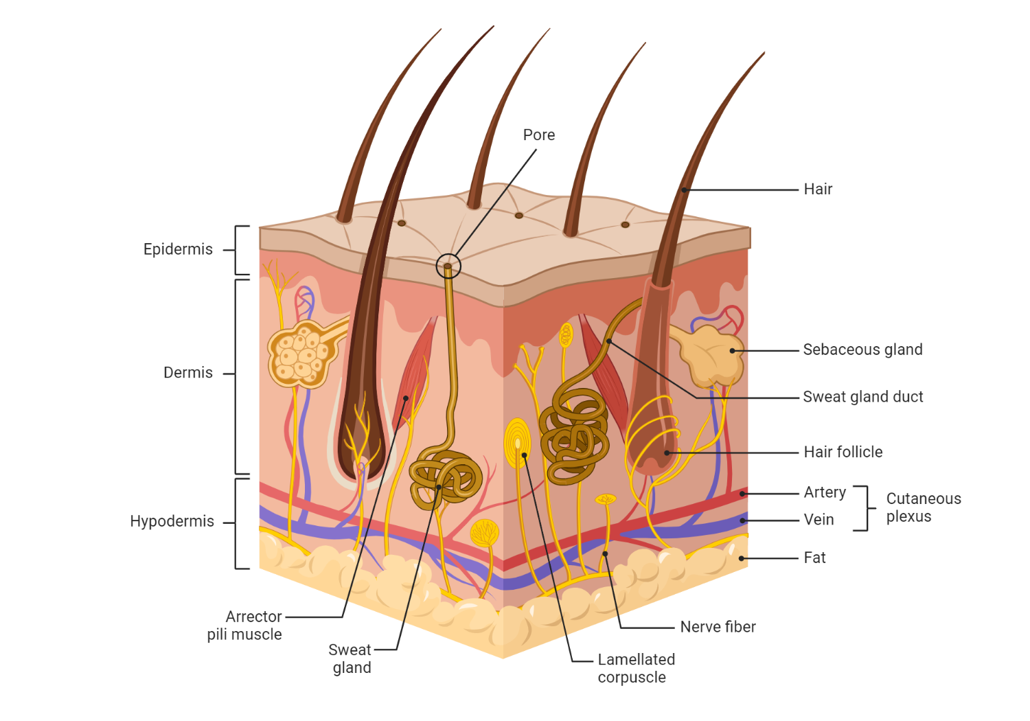
Together, the Basale, Spinosum, and Granulosum form the ‘living layer,’ while the Corneum comprises the ‘dead layer.’ Our skin’s remarkable cell turnover time is typically 4-5 weeks, from the basal layer to desquamation. Additionally, Merkel cells in the basal layer help us sense touch.
Mechanism of action of androgens in the skin
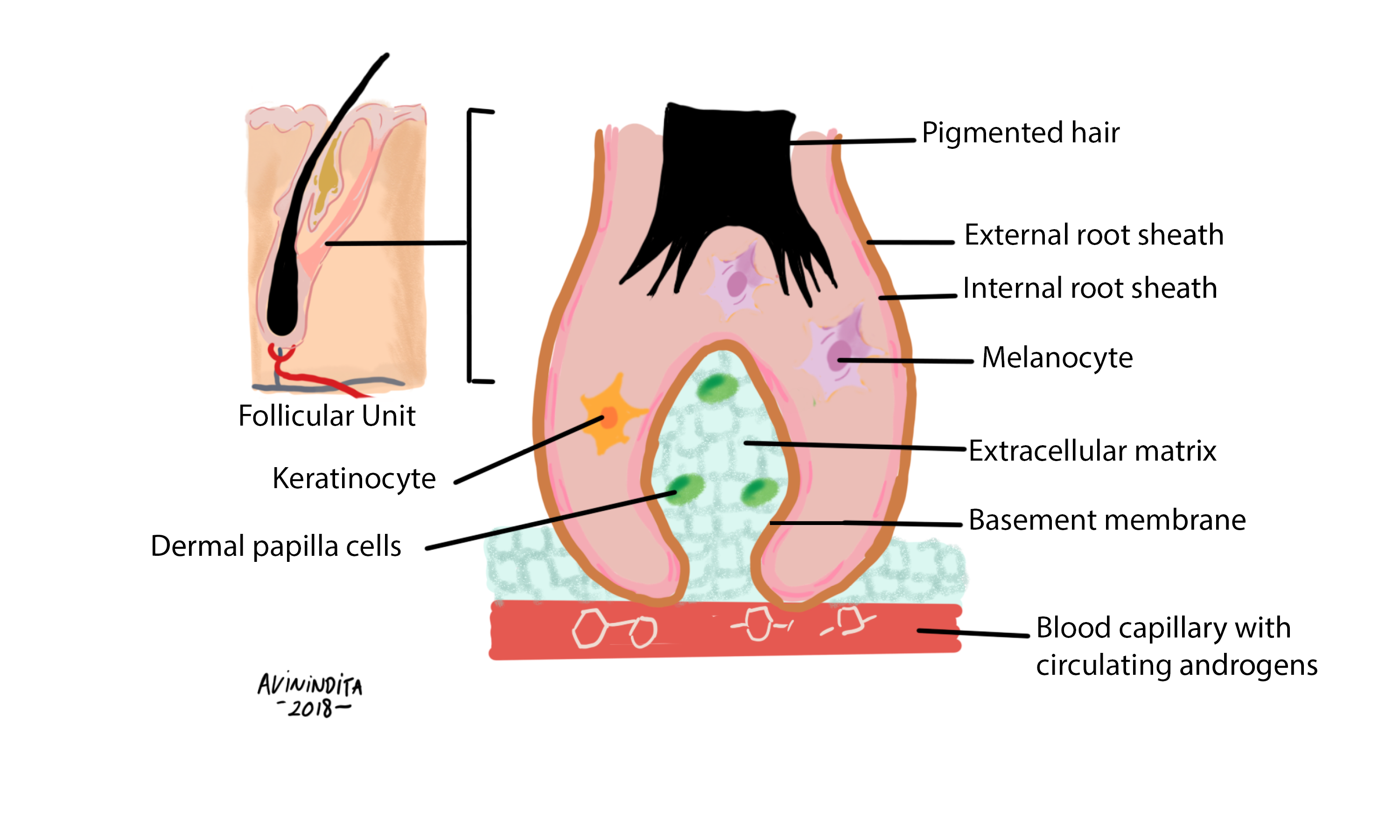
Circulating androgens access the dermal papilla cells via dermal capillaries. Androgens (testosterone or dihydrotestosterone) bind to specific target nuclear receptors in dermal papilla cells. Direct stimulation of keratinocytes and melanocytes occurs through androgen-mediated release of regulatory and growth-promoting factors from the dermal papilla cells.