
The parathyroid glands play a central role in bone and mineral metabolism. Next, the embryologic development, anatomical structure, vascular supply, and functional anatomy will be reviewed.
Historical perspective
The parathyroids were first discovered in an Indian rhinoceros. The first description in humans was reported in a monograph by Ivar Sandstrom (a Swedish medical student) in 1880.
Embryological development of the parathyroid glands
The embryologic structures that lead to the formation of the parathyroids appear around the fifth to sixth weeks of intrauterine life. The primordial tissue that forms the parathyroid glands is formed from the epithelium lining the dorsal portions of the third and fourth pharyngeal pouches. On the other hand, the epithelium lining the ventral portion of the third pharyngeal pouch differentiates into the primordia of the thymus. Two pairs of parathyroid glands are formed and subsequently break off from the third and fourth pharyngeal pouches by the sixth week of gestation.
Parathyroid and thymic primordia from the third pharyngeal pouch migrate in the caudomedial direction. Parathyroid tissue derived from the third pharyngeal pouch comes to a final rest at the caudal level of the thyroid gland (inferior parathyroid glands). Sometimes, parathyroid tissue might descend further with the thymic primordia and may be found close to or within the thymus.
In contrast to the primordia of the third pharyngeal, that of the fourth pharyngeal pouch does not migrate appreciably from its original location and becomes the superior parathyroid glands. The superior parathyroid glands lie at the level of the cricoid cartilage. Variations in the number, size, and locations of the parathyroids can still occur in normal patients.
Anatomical Structure of the Parathyroid gland
The parathyroid glands are ovoid glands that measure 4-6mm x 2-4mm x 0.5-2mm in size. They weigh approximately 30mg in size, with the inferior parathyroids being significantly larger than the superior parathyroids.
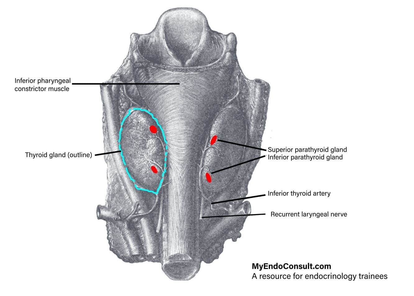 Figure 1. Anatomical relationships of the parathyroid gland
Anatomical Relationships of the Parathyroid gland
The parathyroid glands lie within the cervical fascial sheath of the thyroid gland and are attached to the back of the lateral lobes of the thyroid glands. The superior parathyroid glands are located near the superior pole of the thyroid gland and are lateral to the position of the recurrent laryngeal nerve (as an anatomic reference). The inferior parathyroid glands are located medial to the position of the recurrent laryngeal nerve (as an anatomic reference)
Vascular supply of the parathyroid gland
Arterial anatomy
The inferior thyroid arteries supply both the superior and inferior parathyroid glands (over 90%). Additional tributaries from the other cervical vessels such as the superior thyroid artery, thyroidea ima artery, laryngeal, esophageal, and tracheal vessels supply the parathyroids as well.
Venous anatomy
The parathyroid veins are drained by superior, middle, and inferior thyroid veins. Superior and middle thyroid veins lead into the internal jugular vein and then into the systemic circulation. The inferior thyroid veins drain directly into the brachiocephalic vein.
Clinical Pearl (Functional Anatomy of Endocrine Glands)
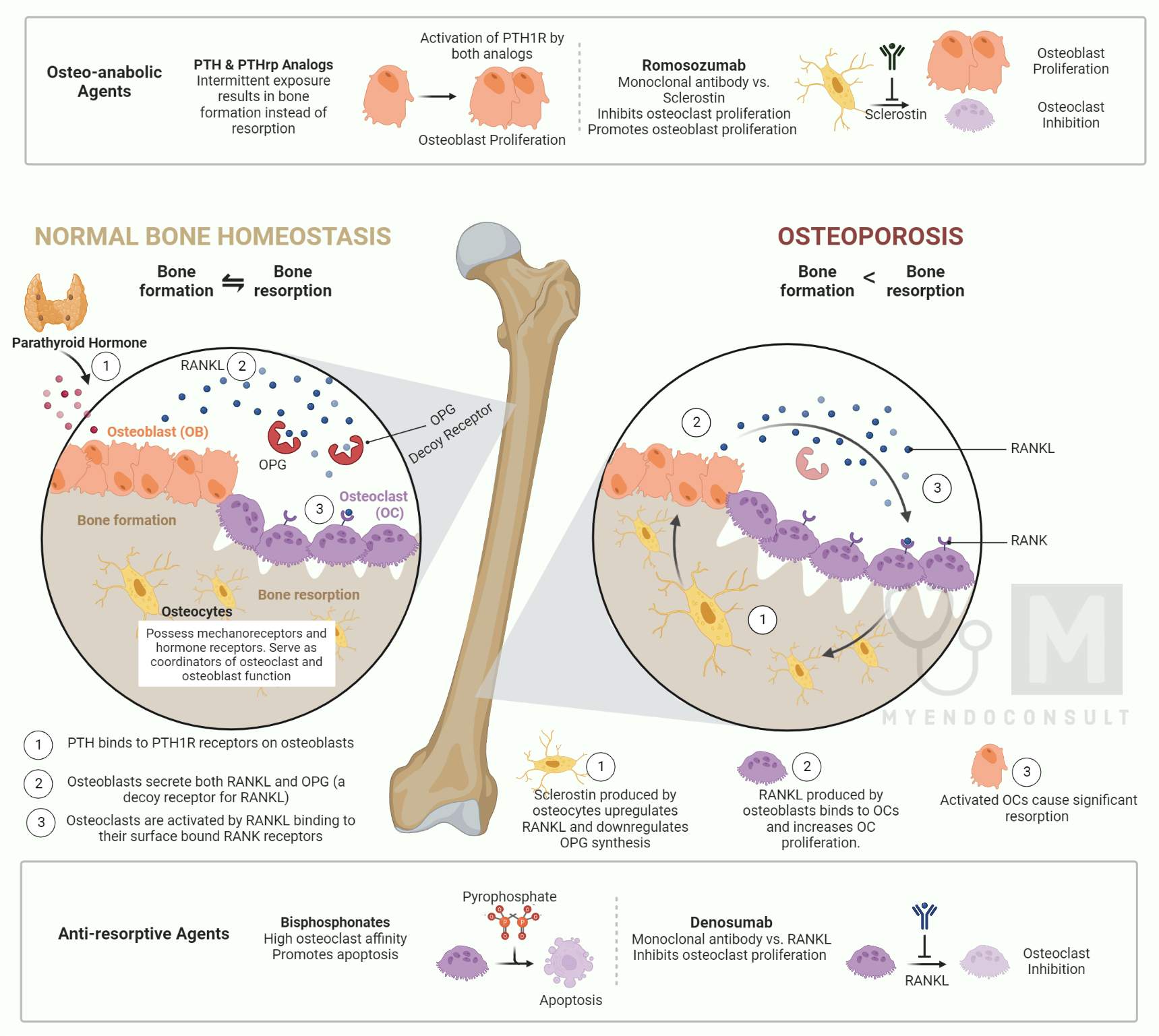
The role of parathyroid hormone in bone resorption
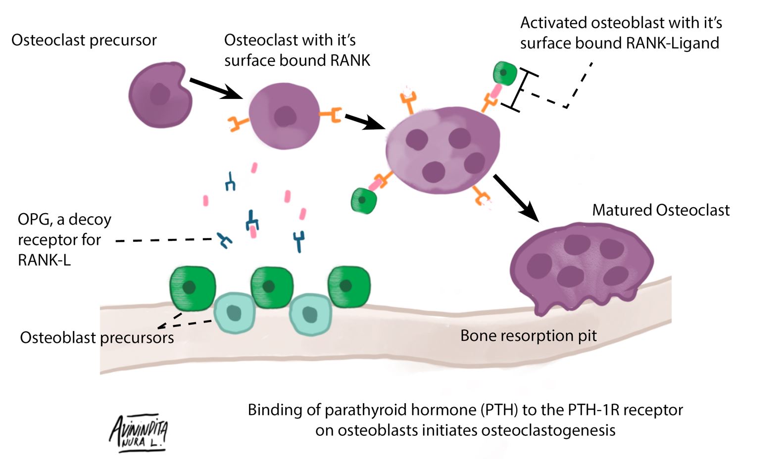 Figure 2. Schematic representation of parathyroid hormone (PTH) mediated osteoblast-osteoclast interaction.
PTH binds to the PTH-1R (Parathyroid hormone 1 receptor) on osteoblasts; this is the first step in the eventual activation of osteoclasts. Osteoblast surface-bound surface-bound receptor activator of nuclear factor κ-B ligand (RANK-L) binds to receptor activator of nuclear factor κ-B (RANK) on osteoclasts leading to differentiation of an osteoclast precursor into a mature osteoclast. Mature osteoclasts present in bone resorption pits are responsible for the liberation of calcium sequestered in hydroxyapatite crystals. Osteoprotegerin (OPG) is a soluble decoy receptor for RANK-L, which provides negative feedback inhibition of osteoclast activation
References
- Hojaij F et al. Parathyroid gland anatomical distribution and relation to anthropometric and demographic parameters: a cadaveric study. Anat Sci Int. 2011 Dec;86(4):204-12.
- Taterra D et al. The prevalence and anatomy of parathyroid glands: a meta-analysis with implications for parathyroid surgery. Langenbecks Arch Surg. 2019 Feb;404(1):63-70.