The pituitary gland (also known as hypophysis) is a pea sized gland that is composed of two significant portions, the anterior and posterior lobes. The adenohypophysis (anterior lobe) and the neurohypophysis (posterior lobe) produce pituitary hormones. The embryology, clinically relevant anatomic relations and vascular supply of the pituitary gland will be reviewed next.
Embryological development of the pituitary gland
The adenohypophysis
The adenohypophysis is a derivative of embryonic oral ectoderm. The neurohypophysis (posterior lobe ) is derived from neural ectoderm located on the floor of the forebrain. During the 4th to 5th week of embryonic development, ectoderm lining the roof of the stomodeum forms a pouchlike recess classically referred to as Rathke’s pouch. This recess extends upwards and touches the forebrain. The surrounding mesoderm then constricts the neck of Rathke’s pouch, which results in a closed cavity surrounded by oral ectoderm.
The remnants of oral ectoderm, which was pinched off by mesoderm during the formation of Rathke’s pouch, is referred to as the “craniopharyngeal canal”. Although this remnant usually involutes during development, it may persist into adult life as a vestigial “pharyngeal pituitary” located on the dorsal wall of the pharynx. More importantly, the pharyngeal pituitary may be a rare source of ectopic pituitary hormonal secretion.
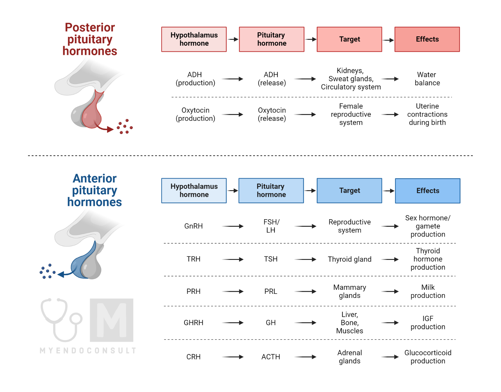
The anterior pituitary produces the following tropic anterior pituitary hormones : Adrenocorticotropic hormone (ACTH), Thyroid stimulating hormone (TSH), follicle stimulating hormone (FSH), luteinizing hormone (LH), Growth hormone (GH), melanocyte stimulating hormone and prolactin.
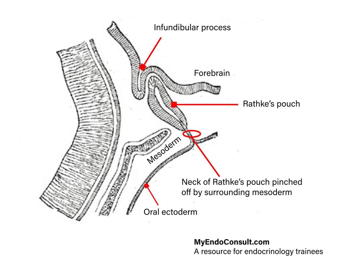 Figure 1. Development of the hypophysis
The neurohypophysis
A neural outgrowth extends downwards from the floor of the third ventricle towards the mouth. This “funnel-shape” outgrowth from the forebrain is the infundibular process. The infundibular process forms the neurohypophysis which is composed of axons and nerve endings of neurons that have their cell bodies in the hypothalamus (supraoptic and paraventricular nuclei). The previously described Rathke’s pouch fuses on each side of the infundibular process and subsequently obliterates its lumen. A remnant of Rathke’s pouch persists and results in the formation of Rathke’s cleft. Colloid-filled cysts may form in this vestigial process and are known as Rathke’s cleft cysts.
The posterior pituitary gland releases antidiuretic hormone (ADH, also known as vasopressin) and oxytocin.
Anatomical Structure of the Pituitary Gland
The anterior lobe (adenohypophysis)
- The pars distalis, also known as pars glandularis form the largest portion of the adenohypophysis.
- The pars tuberalis is a rim of tissue that fuses around the upper portion of the pituitary stalk.
- The pars intermedia is a narrow rim of ectodermal tissue lying next to the neurohypophysis and separated from the pars distalis by the hypophyseal cleft (Rathke’s cleft)
The posterior lobe (neurohypophysis)
- The pars nervosa forms the bulk of the neurohypophysis.
- The median eminence is that part of the tuber cinereum that lies above the pars tuberalis.
- The infundibular stalk (or neural stalk) connects the pars nervosa to the median eminence of the tuber cinereum.
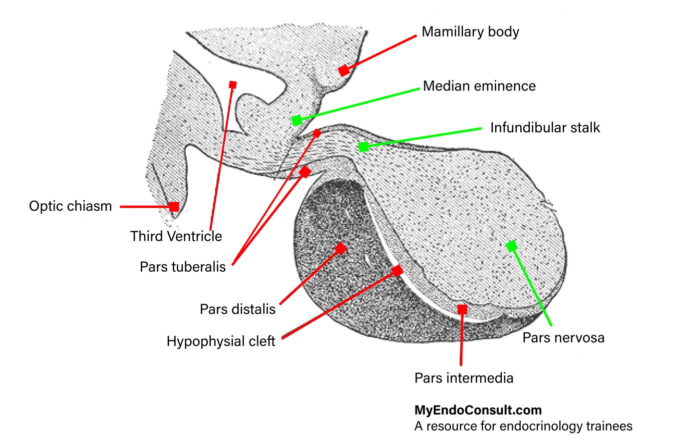 Figure 2. The components of the hypophysis
Anatomical Relationships of the Pituitary Gland
The hypophysis is an ovoid-shaped, reddish-gray gland measuring 12mm x 8mm in its transverse and anterior-posterior dimensions, respectively. The gland measures about 6mm in it’s cranio-caudal distance. The hypophysis sits on the floor of the sellar turcica and is bound on its superior aspect by the optic chiasm. It is bound laterally by the cavernous sinus and anterior-inferiorly by the sphenoid sinus. Finally, the gland is shielded posteriorly by the posterior wall of the sella turcica, pons, and posterior intercaverous sinus.
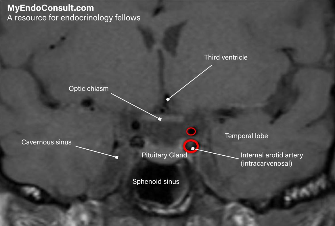 Figure 3. MRI of the pituitary gland (coronal section) showing its relevant anatomical relationships
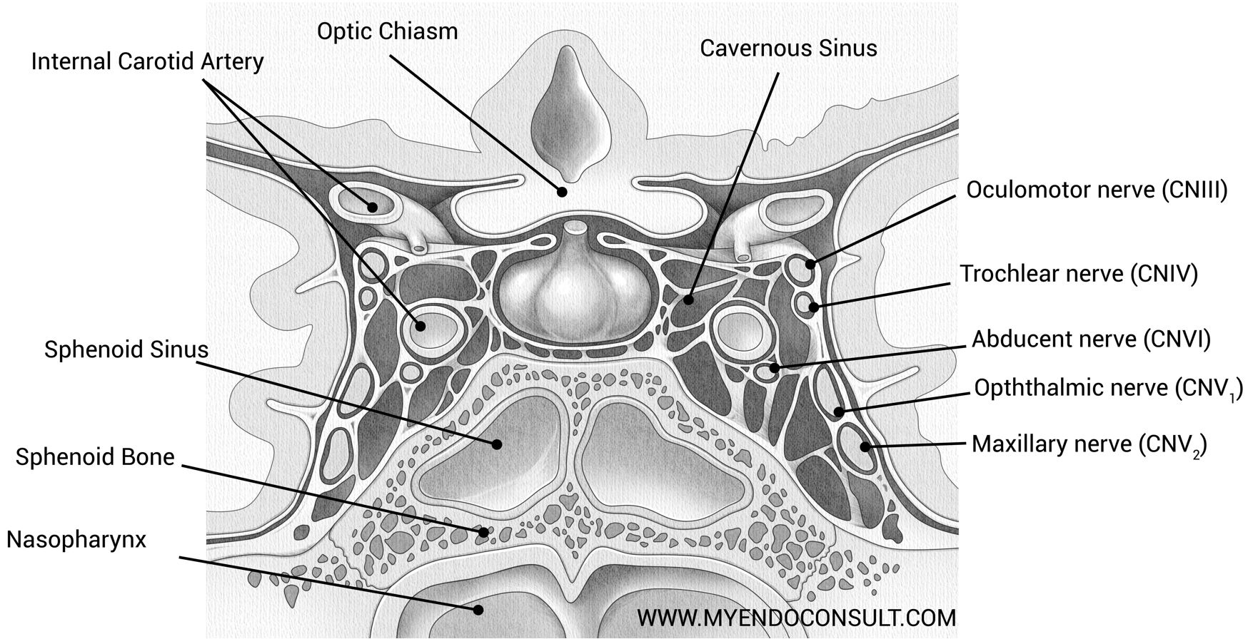 Figure 4.0. The cavernous sinus.
The cavernous sinuses are located on either side of the sphenoid bone and adjacent to the pituitary gland. The medial wall of each cavernous sinus lies the intracavernosal internal carotid artery and abducens nerve (cranial nerve VI). The lateral wall contains the oculomotor (cranial nerve III), trochlear (cranial nerve IV), ophthalmic and maxillary branches of the trigeminal nerve (cranial nerve V).
Vascular supply of the pituitary gland
Arterial anatomy
The pituitary gland is supplied by a pair of arteries – the right and left superior hypophysial arteries from above and the right and left inferior hypophysial arteries from below. Each superior hypophysial artery bifurcates into anterior and posterior branches, which permeate the pituitary stalk. The inferior hypophysial arteries arise from the intracavernous segment of each internal carotid artery. Branches of the inferior and superior hypophysial arteries anastomose freely within the pituitary gland.
Venous anatomy
It is worth noting that epithelial tissue in the pars distalis of the adenohypophysis does not receive direct arterial blood supply from either the superior or inferior hypophysial arteries. The adenohypophysis receives its blood supply from hypophysial portal vessels which are supplied by capillary vessels within the median eminence and the infundibular stem. The adenohypophysis is drained by venous dural sinuses, which surround the pituitary and subsequently lead to systemic circulation.
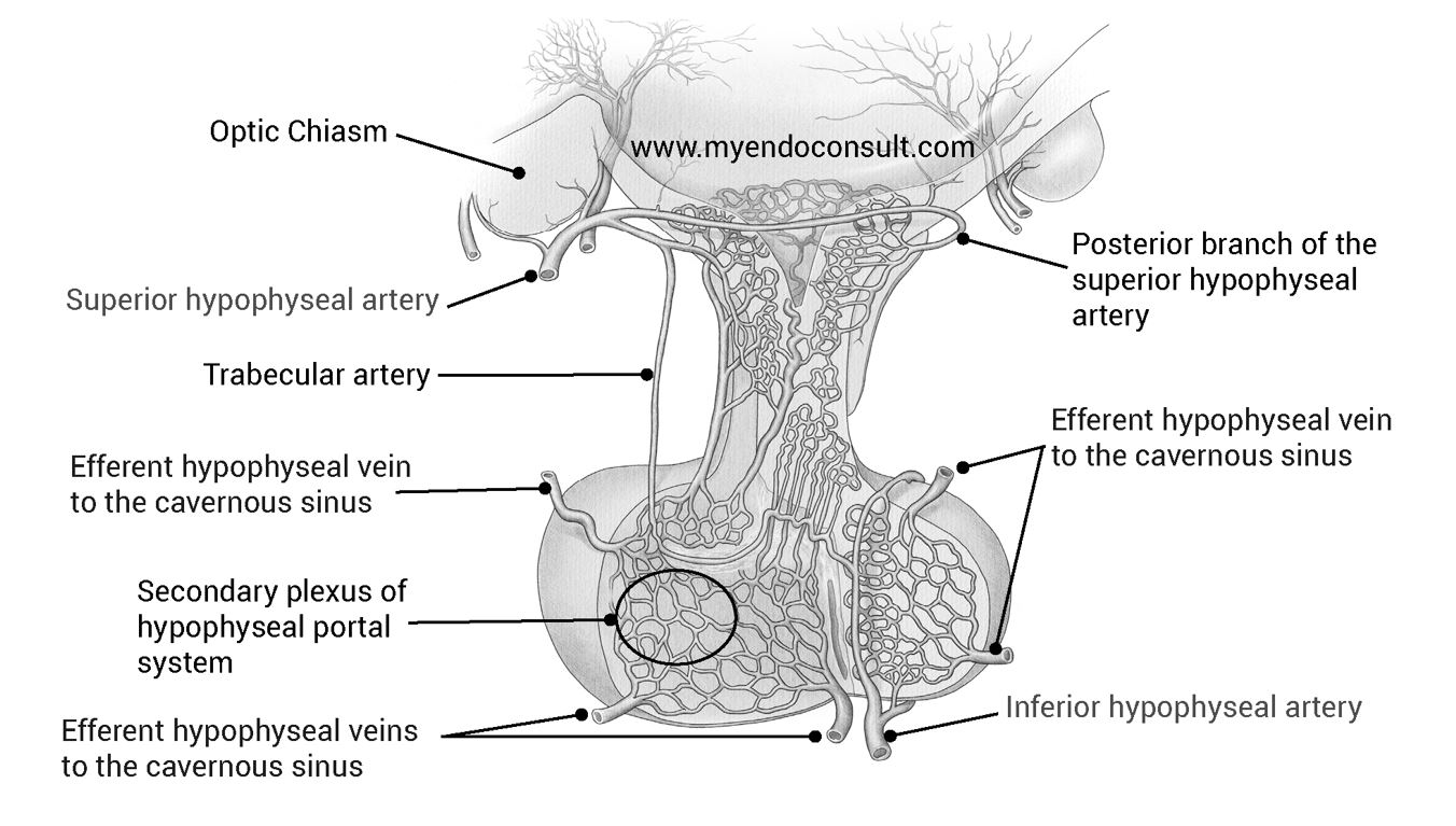 Figure 5. Venous and arterial supply of the pituitary gland. AH adenohypophysis, NH neurohypophysis
A schematic representation of the pituitary gland showing relevant arterial (superior hypophyseal artery and inferior hypophyseal artery) and venous supply (multiple efferent hypophyseal veins draining into the cavernous sinus)
What about the vascular supply of the anterior pituitary gland predisposes it to infarction.
The hypophysis is drained mainly by portal vessels. Thus, extensive occlusion of this venous network can lead to ischemic necrosis of the adenohypophysis since these vessels are the only afferent supply to the sinusoids of the pars distalis (majority of the anterior pituitary). Also, read this article about pituitary apoplexy.
References
- Amar AP, Weiss MH. Pituitary anatomy and physiology. Neurosurg Clin N Am. 2003 Jan;14(1):11-23, v.
- Daniel PM. Anatomy of the hypothalamus and pituitary gland. J Clin Pathol Suppl (Assoc Clin Pathol). 1976;7:1-7.