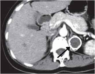
For adrenal adenomas with concerning computed tomographic features, adrenal washout characteristics can be used to characterize these lesions further. By administering intravenous contrast, the contrast uptake phase can be compared to the washout phase in order to differentiate between benign and malignant adrenal lesions. For non-contrast CT of the adrenal glands, an attenuation value less than 10 Hounsfield Units (HU) has a high specificity for lipid-rich adrenal adenomas.
You will need the following parameters in order to calculate the washout characteristics of an adrenal nodule.
- HU (portal venous phase) = HU estimation during the portal venous phase after contrast administration
- HU (delayed phase) = HU estimation of the adrenal adenoma, approximately 15 minutes after contrast administration (delayed)
- HU (pre-contrast phase) = HU estimation of the adrenal adenoma before administration of IV contrast
Absolute washout:
[(HUportal venous phase) – (HUdelayed phase)] / [(HUportal venous phase) – (HUpre-contrast phase)] x 100
Relative washout:
[(HUportal venous phase) – (HUdelayed phase)] / (HUportal venous phase) x 100
Interpretation of adrenal washout
- An absolute washout >60% is suggestive of an adrenal adenoma
- A relative washout >40% is suggestive of an adrenal adenoma
- Limited washout on the other hand may be due to either pheochromocytomas or adrenocortical carcinoma
Origin of Computed Tomography
Godfrey Hounsfield was the first to recognize that x-ray measurements taken from different directions could lead to a reasonably accurate reconstruction of internal tissues. Hounsfield showed that a computation of the x-ray attenuation coefficient in each CT slice area could be used to image tissues of varying densities in the human body. Hounsfield won the 1979 Nobel Prize in Physiology and Medicine for his pioneering work in computed tomography.
Estimating Hounsfield Units on CT
Use the region of interest tool to measure the HU (see the illustration below).

References
- Hounsfield GN. Computerized transverse axial scanning (tomography): part 1. Description of system. Brit J Radiol. 1973;46:1016–22.
- Song JH, Chaudhry FS, Mayo-Smith WW. The incidental adrenal mass on CT: prevalence of adrenal disease in 1,049 consecutive adrenal masses in patients with no known malignancy. AJR Am J Roentgenol. 2008;190(5):1163–8.
Kindly Let Us Know If This Was helpful? Thank You!


