

The Hounsfield unit (HU) measures the attenuation of X-rays by tissue. It is defined as the ratio of the intensity of the X-rays beams after passing through the tissue to the intensity of the incident beam. The unit is named after Sir Godfrey Newbold Hounsfield, who co-invented computed tomography (CT) scanning.
Hounsfield unit scale and chart
The HU can be used to quantify different tissues on a CT scan. For example, water has a HU of 0, fat has a HU of -100 to -150, and bone has a HU of +1000. Tumors also have characteristic HUs that can be used to distinguish them from normal tissues. The table below depicts the Hounsfield unit values of various tissues on multidetector CT imaging.
| Substance | HU |
| Air | -1000 |
| Lung | -500 |
| Fat | -100 to -50 |
| Water | 0 |
| Cerebrospinal fluid | 15 |
| Renal Parenchyma | 30 |
| Blood | +30 to +45 |
| Muscle | +10 to +40 |
| Brain (Grey matter) | +37 to +45 |
| Brain (White matter) | +20 to +30 |
| Liver | +40 to +60 |
| Soft tissue, IV Contrast | +100 to +300 |
| Trabecular bone | +700 |
| Cortical Bone | +2000 to +3000 |
Application of Hounsfield unit in clinical practice
The HU scale is a measure of the attenuation of x-rays by tissues. It is used extensively in diagnostic radiology, particularly for CT scans. The HU scale ranges from -1000 to +1000, with water having a Hounsfield unit of 0.
Tissues with a high HU (such as bone) absorb more x-rays than tissues with a low HU (such as lung tissue). The application of HU allows radiologists to more accurately interpret CT images. For example, a high HU indicates the presence of dense tissue, which may be suggestive of pathology. By contrast, a low HU indicates the presence of less dense tissue, which is typically seen in normal organs. The use of CT attenuation value of tissues has helped to improve the accuracy of diagnosis and has led to better patient care.
The Hounsfield unit can be useful in both clinical and diagnostic research. For example, it can be used in determining the severity of diseases such as pneumonitis and pulmonary edema. It can also help to identify areas of necrosis in tumors. In addition, the HU can be used to monitor the progression of diseases such as cancer, since changes in the attenuation of X-rays by tissue can indicate changes in the disease state.
Application of HU in clinical endocrinology
An adrenal incidentaloma is defined as a mass >1cm diameter discovered incidentally in radiology studies. These lesions should be evaluated to determine their functional and malignant status.
The determination of whether an adrenal incidentaloma is benign or malignant is based on its size, attenuation by measuring HU (pre-contrast phase), and time of contrast washout (post-contrast phase). Estimation of an adrenal incidentaloma’s attenuation on a non-contrast CT scan is compared relative to that of fat tissue (-20 to -150) and the kidney (20 to 150HU).
The functional status of an adrenal incidentaloma is dependent on hormonal assays (Review the dexamethasone suppression test here)
Functional adrenal masses (pheochromocytomas, aldosteronomas, and cortisol-producing tumors) should be referred for surgical resection. For non-functioning adrenal masses which appear benign based on computed tomography attenuation value, a repeat scan in 6-12 months is reasonable.
All patients diagnosed with an adrenal incidentaloma, require a repeat CT scan of the adrenal glands at 6, 12, and 24 months respectively. Earlier scans may be required for suspicious nodules. It is worth noting that pheochromocytomas typically grow at a rate of 1.0 cm per year, while adrenocortical carcinomas have a growth rate of 2.0 cm per year. Use this calculator to determine the washout characteristics of an adrenal nodule.
Benign findings of an adenoma
- Low attenuation (<10 Hounsfield units) and <4cm
- Smooth border with sharp margins
- Homogenous
- Unilateral
- Rapid contrast washout, >50% after 10minutes of contrast administration.
A very low CT attenuation value (–10 to –20HU) is characteristic of adrenal myelolipoma.
Malignant findings of an adenoma
- High attenuation (>20 Hounsfield units) and >4cm diameter
- Increased mass vascularity
- Delayed contrast medium washout, <50% after 10 minutes of contrast administration.
- May be unilateral or bilateral.
- Presence of necrosis, hemorrhage, or calcification
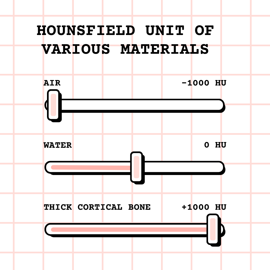
Visual aid for remembering the attenuation value (Hounsfield units) of various materials under cone beam computed tomography.
References
- Zeiger M, Thompson G, Duh QY, Hamrahian A, Angelos P, Elaraj D, Fishman E, Kharlip J. American Association of Clinical Endocrinologists and American Association of Endocrine Surgeons medical guidelines for the management of adrenal incidentalomas. Endocr Pract. 2009;15 Suppl 1:450–3.
- Lim Fat, Daren; Kennedy, Jim; Galvin, Rose; O’Brien, Fergal; Mc Grath, Frank; Mullett, Hannan (2012-05-01). “The Hounsfield value for cortical bone geometry in the proximal humerus—an in vitro study”. Skeletal Radiology. 41 (5): 557–568.