Bone age assessment is an essential diagnostic tool used by physicians to evaluate skeletal development and diagnose growth-related conditions. It involves estimating an individual’s bones’ maturity based on their radiographic appearance. This information can be used to predict future growth, identify any developmental abnormalities, and monitor the effectiveness of treatments.
The most commonly used method for bone age assessment is the Greulich and Pyle method, which involves comparing the radiographic appearance of an individual’s left hand and wrist bones to a standardized atlas of bone development.
The atlas consists of a series of radiographs of the left hand and wrist bones of healthy children of various ages and sexes. The physician can determine the individual’s bone age by comparing an individual’s radiographs to the atlas.
Greulich and Pyle method
The process of bone age assessment involves several steps:
Obtain Radiographs:
The first step is to obtain radiographs of the individual’s left hand and wrist bones. The radiographs should be taken using standardized techniques to ensure accurate results.
Identify Bones:
The physician must identify the bones visible in the radiographs. The bones that are used for bone age assessment include the radius, ulna, carpals, and phalanges.
Compare to Atlas:
The physician then compares the radiographic appearance of the individual’s bones to the corresponding images in the Greulich and Pyle atlas. The physician should consider both the size and shape of the bones when making the comparison.
Determine Bone Age:
Once the physician has compared the individual’s bones to the atlas, they can estimate the individual’s bone age. The bone age is usually reported in months or years and should be interpreted in the context of the individual’s chronological age and other relevant clinical information.
It is important to note that bone age assessment in general is not a perfect method for evaluating skeletal development. There is a degree of variation in bone development between individuals, and bone age assessment may not accurately predict future growth. Additionally, the Greulich and Pyle atlas may not be applicable to all populations, and physicians should consider using alternative methods for bone age assessment in some cases.
Tanner-Whitehouse method
The Tanner-Whitehouse method, also known as the TW2 method, is a system for assessing bone age in children that takes into account the size and shape of individual bones as well as other physical characteristics of the child. It was first introduced in the 1960s as an improvement over the Greulich and Pyle method, which had limitations in terms of its applicability to children of different ethnicities and body types.
The TW2 method involves evaluating the appearance of 20 bones in the hand and wrist, as well as the radius and ulna in the forearm.
Each bone is assigned a score based on its size and shape, and these scores are then combined to give an overall assessment of bone age. The method also takes into account other physical characteristics of the child, such as height, weight, and sexual development.
The TW2 method has been found to be more accurate than the Greulich and Pyle method in assessing bone age in children with conditions that affect growth and development, such as Turner syndrome and congenital adrenal hyperplasia. It is also considered to be more reliable than other methods that rely solely on a radiographic assessment of bone development.
One potential drawback of the TW2 method is that it can be time-consuming and requires specialized training to perform accurately. Additionally, like any method of bone age assessment, there is some degree of variability and potential for error. Therefore, it is important for physicians to consider the limitations of the TW2 method and to interpret results in the context of other clinical information.
In summary, the Tanner-Whitehouse method is a system for assessing bone age in children that takes into account the size and shape of individual bones as well as other physical characteristics of the child. It is considered to be a more accurate and reliable method than the Greulich and Pyle method in certain clinical settings, but requires specialized training and should be used in combination with other clinical information to make an accurate diagnosis
Computer-generated (Artificial Intelligence) Assessment of Bone Age
Computer-generated assessment of bone age is a relatively new and promising method for assessing skeletal maturity in children. This approach involves using computer algorithms to analyze radiographs of the hand and wrist and automatically calculate a bone age score based on the appearance of the bones.
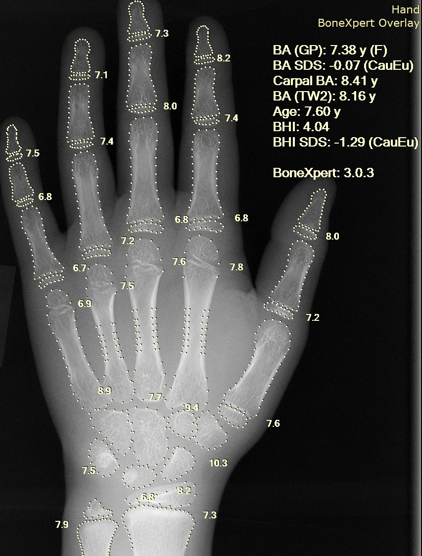
Source of image : WikiCommons
One advantage of computer-generated bone age assessment is that it eliminates the need for a trained radiologist or physician to interpret the radiographs, which can save time and reduce the potential for human error. The use of computer algorithms can also help to standardize bone age assessments across different institutions and eliminate inter-observer variability.
The process of computer-generated bone age assessment typically involves using software that has been trained on large datasets of radiographs and corresponding bone age scores.
The software analyzes the radiographs to identify and measure specific bone features, such as the length and shape of the bones, and then applies a machine learning algorithm to predict the most likely bone age score based on these measurements.
Studies have shown that computer-generated bone age assessment can be as accurate as or more accurate than traditional methods of bone age assessment, such as the Greulich and Pyle method or the Tanner-Whitehouse method. This technology has the potential to improve the speed and accuracy of bone age assessment, particularly in settings where there may be limited access to specialized radiologists or pediatric endocrinologists.
However, it is important to note that computer-generated bone age assessment is not without limitations. The accuracy of the assessment depends on the quality of the radiographs and the algorithm used, and there may be variability in bone development within the same age group that can affect the accuracy of the prediction. Additionally, the use of computer algorithms cannot replace the importance of clinical judgment and other clinical information in making a diagnosis.
In summary, computer-generated assessment of bone age is a promising new method for evaluating skeletal maturity in children. It has the potential to improve the accuracy and standardization of bone age assessment, particularly in settings where there may be limited access to specialized radiologists or pediatric endocrinologists. However, like any method of bone age assessment, it should be used in combination with other clinical information to make an accurate diagnosis.
References
Greulich WW, Idell PS. Radiographic atlas of skeletal development of the hand and wrist. Am J Med Sci. 1959;238:393.
Tanner JM, Whitehouse RH, Cameron N. Assessment of skeletal maturity and prediction of adult height (Tw2 method). 1989.
Lee H, Tajmir S, Lee J, Zissen M, Yeshiwas BA, Alkasab TK, Choy G, Do S. Fully Automated Deep Learning System for Bone Age Assessment. J Digit Imaging. 2017 Aug;30(4):427-441. doi: 10.1007/s10278-017-9955-8. PMID: 28275919; PMCID: PMC5537090.
Li, S., Liu, B., Li, S. et al. A deep learning-based computer-aided diagnosis method of X-ray images for bone age assessment. Complex Intell. Syst. 8, 1929–1939 (2022).