Hashimoto's thyroiditis, also known as chronic lymphocytic thyroiditis, is an autoimmune disorder that results in inflammation and damage to the thyroid gland. It is the most common cause of hypothyroidism, which is characterized by insufficient production of thyroid hormones. In this review, we will provide a detailed description of the histological features of Hashimoto's thyroiditis.
Histology of Hashimoto's Thyroiditis

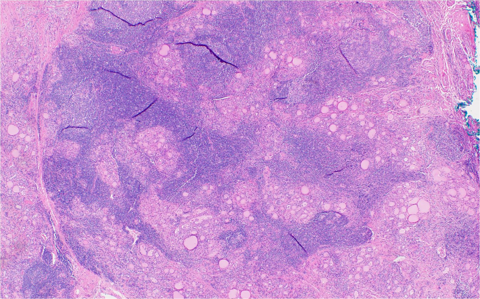
Hashimoto’s thyroiditis characterized by dense lymphoplasmacytic infiltrate of thyroid parenchyma
Parenchymal destruction: The thyroid parenchyma in Hashimoto's thyroiditis is characterized by extensive infiltration of lymphocytes, leading to the destruction of thyroid follicles and replacement of the normal architecture by fibrosis.
Lymphocytic infiltration: One of the key histological findings of Hashimoto's thyroiditis is the infiltration of the thyroid gland by lymphocytes.
These lymphocytes form aggregates, leading to the development of lymphoid follicles with germinal centers, which are a hallmark of the autoimmune response in this condition.
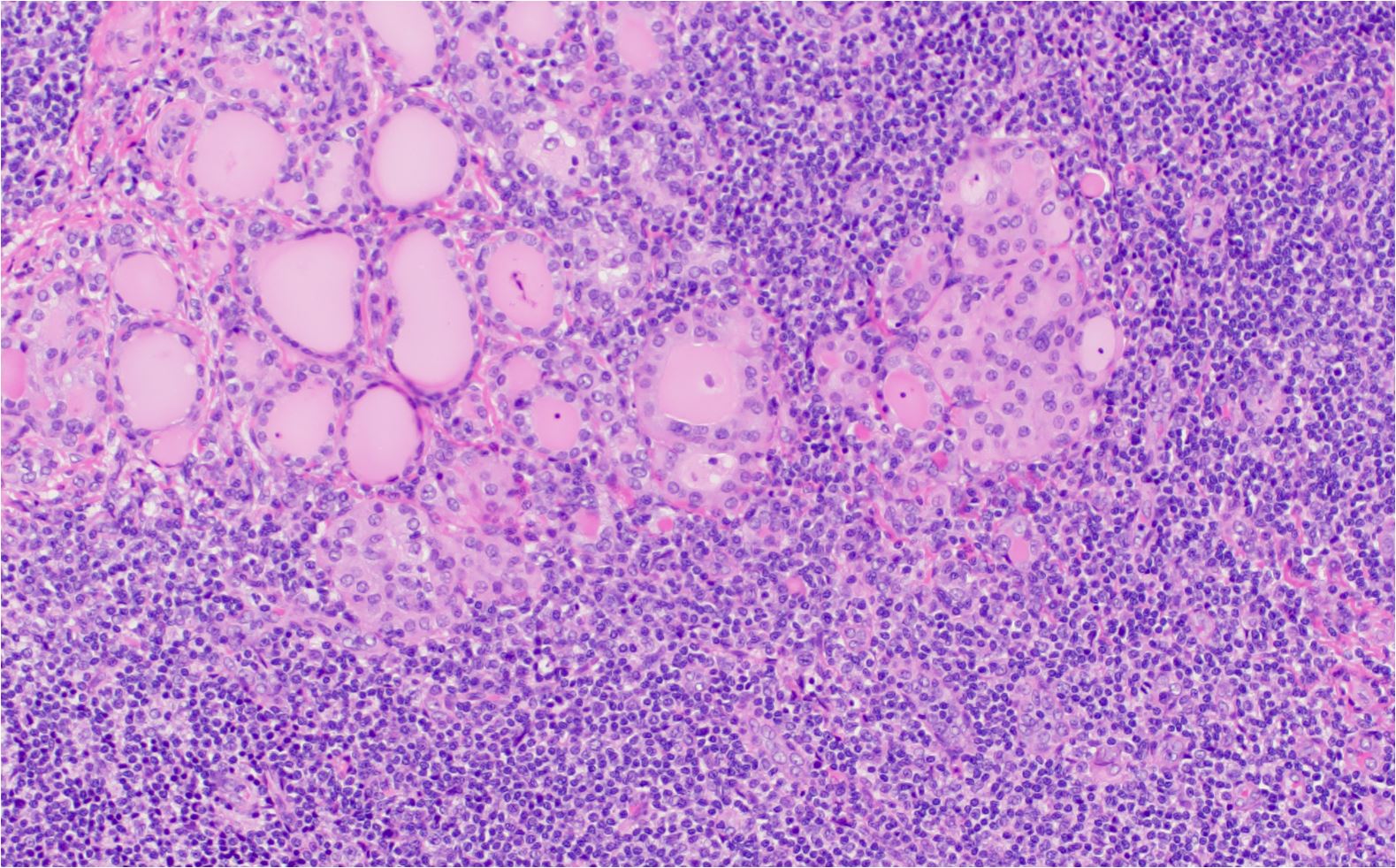
Hurthle cells (oxyphilic cells): In Hashimoto's thyroiditis, the follicular epithelial cells may undergo a metaplastic change, transforming into large eosinophilic cells known as Hurthle cells or oxyphilic cells.
Also, these cells have abundant granular, eosinophilic cytoplasm due to the presence of numerous mitochondria. Furthermore, the nuclei of Hurthle cells are large, round, and hyperchromatic with prominent nucleoli.
Atrophy and fibrosis: As the disease progresses, the destruction of thyroid follicles and the parenchyma is accompanied by increasing fibrosis, leading to the atrophy of the thyroid gland. Indeed, the fibrosis may be patchy or diffuse, depending on the stage and severity of the disease.
Follicular cell degeneration: The thyroid follicular cells in Hashimoto's thyroiditis may exhibit degenerative changes, such as apoptosis or necrosis, due to the autoimmune-mediated destruction of the gland.
Variable colloid content: The thyroid follicles in Hashimoto's thyroiditis often contain variable amounts of colloid, which may be scant, patchy, or even absent in some cases. This irregularity is due to the destruction of the follicular architecture and impaired hormone synthesis.
Multinucleated giant cells: Occasionally, multinucleated giant cells may be present in Hashimoto's thyroiditis, resulting from the fusion of macrophages as they attempt to phagocytose the degenerating thyroid follicular cells.
H and E features of Hashimoto's Thyroiditis

Hashimoto’s thyroiditis characterized by dense lymphoplasmacytic infiltrate and Hurthle cell metaplasia. Large areas of thyroid parenchyma are replaced by inflammatory infiltrate
In summary, the histological attributes of Hashimoto's thyroiditis include parenchymal destruction, lymphocytic infiltration with the formation of lymphoid follicles and germinal centers, the presence of Hurthle cells, atrophy and fibrosis of the thyroid gland, follicular cell degeneration, variable colloid content, and occasional multinucleated giant cells.
These features are indicative of the autoimmune pathogenesis of the condition and help distinguish Hashimoto's thyroiditis from other thyroid disorders.
Kindly Let Us Know If This Was helpful? Thank You!


