Hurthle Cell Adenoma
Hurthle cell adenoma is a rare, benign tumor of the thyroid gland that originates from the follicular cells. Hurthle cells, also known as oxyphilic or oncocytes, are characterized by their abundant granular, eosinophilic cytoplasm, and large hyperchromatic nuclei. In this review, we will provide a detailed description of the histological features of Hurthle cell adenoma.
Histological features of Hurthle Cell Adenoma

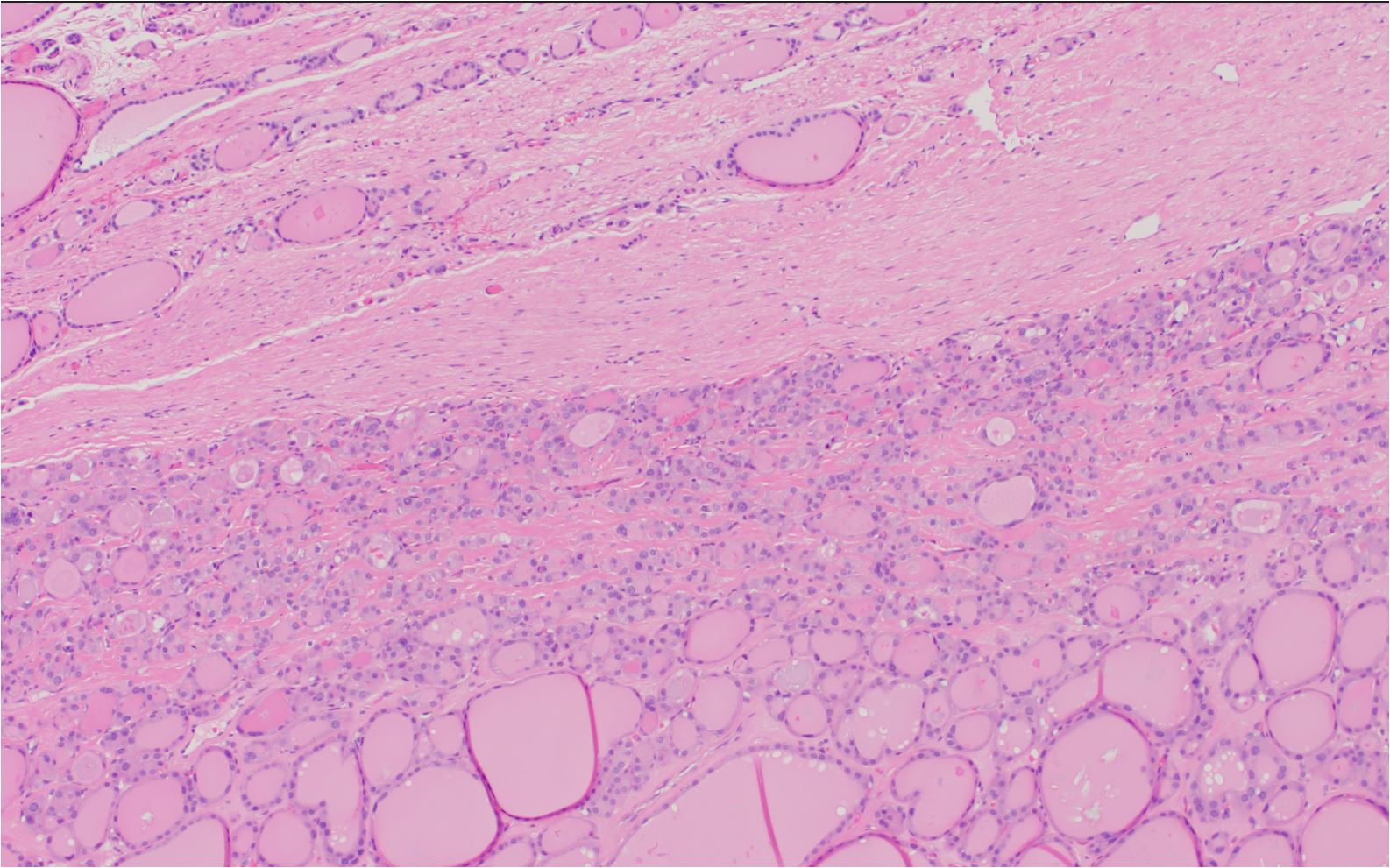
Hurthle cell adenoma with thick fibrous capsule. No capsular or vascular invasion could be identified
Gross appearance: Hurthle cell adenomas typically present as well-circumscribed, solitary, round or oval nodules within the thyroid gland. The size of the tumors may vary, but they are usually a few centimeters in diameter. Also, the cut surface of the tumor may appear solid, firm, and tan to reddish-brown in color.
Encapsulation: Hurthle cell adenomas are usually encapsulated by a thin, fibrous capsule that separates the tumor from the adjacent thyroid parenchyma.
The presence of encapsulation distinguishes Hurthle cell adenomas from other thyroid neoplasms and benign lesions, such as nodular hyperplasia.
Cellular architecture: The architecture of Hurthle cell adenomas is predominantly solid or trabecular, with tumor cells arranged in sheets, cords, or nests. The tumor cells may occasionally form microfollicles, but these structures are less common than in other follicular neoplasms.
Hurthle cells: The defining characteristic of Hurthle cell adenoma is the presence of Hurthle cells. These cells are large, polygonal cells with abundant granular, eosinophilic cytoplasm due to the presence of numerous mitochondria.
The nuclei of Hurthle cells are large, round, and hyperchromatic, with prominent nucleoli. They typically have well-defined cell borders and form tight intercellular junctions.
Nuclei: The nuclei of Hurthle cells in adenomas are usually uniform in size and shape, with evenly distributed chromatin. Nuclear pleomorphism and atypia are generally minimal or absent in benign Hurthle cell adenomas.
Colloid: Unlike typical follicular adenomas, Hurthle cell adenomas have scant to absent colloid content. This attribute is due to the solid or trabecular arrangement of tumor cells, which do not form well-developed follicular structures.
Mitotic activity: The mitotic rate in Hurthle cell adenomas is usually low, reflecting the benign nature of these tumors.
High mitotic activity may raise suspicion for a malignant Hurthle cell neoplasm, such as Hurthle cell carcinoma.
Capsular and vascular invasion: Hurthle cell adenomas typically lack capsular or vascular invasion, which differentiates them from Hurthle cell carcinomas. The absence of these findings is indicative of a more indolent, benign course.
In summary, the histological features of Hurthle cell adenoma include well-circumscribed, encapsulated nodules, a predominantly solid or trabecular architecture, the presence of characteristic Hurthle cells with abundant eosinophilic cytoplasm and large hyperchromatic nuclei, scant to absent colloid content, low mitotic activity, and the absence of capsular or vascular invasion. These histologic attributes help to distinguish Hurthle cell adenomas from other thyroid lesions and malignant Hurthle cell neoplasms.
Hurthle Cell Carcinoma
Hurthle cell carcinoma, also known as oxyphilic or oncocytic carcinoma, is a rare and distinct subtype of follicular thyroid carcinoma. It arises from Hurthle cells (oxyphilic cells) and has unique histological features. In this review, we will provide a detailed description of the histological features of Hurthle cell carcinoma.
Histological features of Hurthle Cell Carcinoma

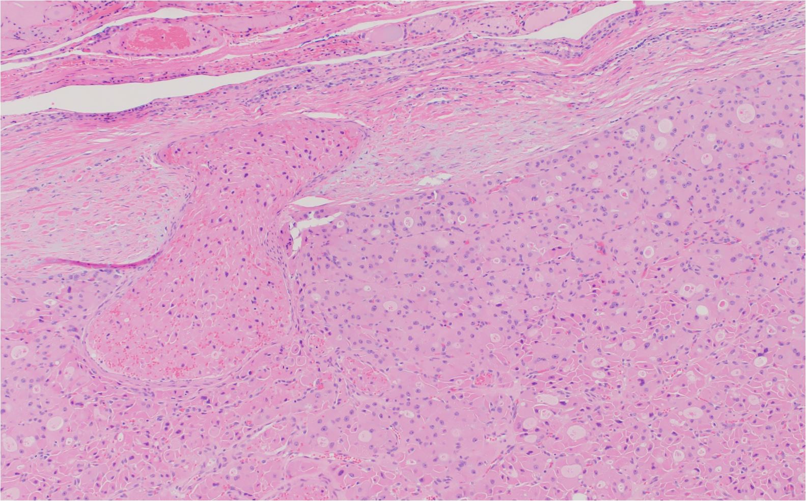
Hurthle cell carcinoma with a focus of vascular invasion into a thick capsule
Gross appearance: Hurthle cell carcinomas typically present as solitary, round or oval nodules within the thyroid gland. They can vary in size but are often larger than benign Hurthle cell adenomas. The cut surface of the tumor may appear solid, firm, and tan to reddish-brown in color.
Encapsulation: Hurthle cell carcinomas are usually well-circumscribed and may be partially or completely encapsulated by a fibrous capsule. However, unlike Hurthle cell adenomas, carcinomas may exhibit capsular and vascular invasion, which are characteric findings indicative of malignancy.
Cellular architecture: The architecture of Hurthle cell carcinoma is predominantly solid, trabecular, or insular, with tumor cells arranged in sheets, cords, or nests. The tumor cells may occasionally form microfollicles, but these structures are less common than in other follicular neoplasms.
Hurthle cells: The defining histologic attribute of Hurthle cell carcinoma is the presence of Hurthle cells. These cells are large, polygonal cells with abundant granular, eosinophilic cytoplasm due to the presence of numerous mitochondria.
The nuclei of Hurthle cells are large, round, and hyperchromatic, with prominent nucleoli. They typically have well-defined cell borders and form tight intercellular junctions.
Nuclei: In contrast to Hurthle cell adenomas, the nuclei of Hurthle cells in carcinomas may exhibit more nuclear pleomorphism, hyperchromasia, and irregularity. These findings can raise suspicion for malignancy.
Colloid: Hurthle cell carcinomas, like Hurthle cell adenomas, have scant to absent colloid content. Furthermore, the presence of solid or trabecular arrangement of tumor cells, results in the formation of poorly-developed follicular structures.
Mitotic activity: The mitotic rate in Hurthle cell carcinomas may be higher than in benign Hurthle cell adenomas. Increased mitotic activity can be a sign of malignancy and aggressive behavior.
Capsular and vascular invasion: A key diagnostic feature of Hurthle cell carcinoma is the presence of capsular and/or vascular invasion. Capsular invasion refers to the infiltration of tumor cells through the capsule that surrounds the tumor mass.
Vascular invasion is identified by the presence of tumor cells within the endothelium-lined spaces of blood vessels within or immediately adjacent to the tumor capsule.
These attributes distinguish Hurthle cell carcinoma from benign Hurthle cell adenoma and are associated with a higher risk of metastasis and recurrence.
Lymph node and distant metastasis: Hurthle cell carcinomas have a higher propensity for lymph node and distant metastasis compared to follicular carcinomas. The most common sites for metastasis are regional lymph nodes, lungs, and bones.
In summary, the histological features of Hurthle cell carcinoma include well-circumscribed nodules that may be partially or completely encapsulated, a predominantly solid or trabecular architecture, the presence of characteristic Hurthle cells with abundant eosinophilic cytoplasm and large hyperchromatic nuclei, scant to absent colloid content, increased mitotic activity, and the presence of capsular and/or vascular invasion. These findings help to distinguish Hurthle cell carcinoma from benign Hurthle cell adenomas
Kindly Let Us Know If This Was helpful? Thank You!


