Normal thyroid follicular cells, also known as thyrocytes, are the primary cell type in the thyroid gland responsible for the production and secretion of thyroid hormones.
These hormones, thyroxine (T4) and triiodothyronine (T3), play crucial roles in regulating metabolism, growth, and development. In this review, we will provide a detailed description of the histological features of normal thyroid follicular cells.
Histological Features of Thyroid Follicular Cells

- Morphology: Thyroid follicular cells are cuboidal or low columnar in shape, depending on their functional state. In an active metabolic state, they are taller and more columnar, while in a resting state, they are shorter and more cuboidal.
- Location: These cells line the walls of the thyroid follicles, which are spherical structures filled with colloid. The colloid serves as a reservoir for the storage of thyroid hormones in the form of thyroglobulin, a large glycoprotein.
- Nucleus: Thyroid follicular cells have a central, round to oval-shaped nucleus with a visible nucleolus. The chromatin is evenly distributed, and the nuclear membrane is well-defined, with the nucleus situated basally located within the cell.
- Cytoplasm: The cytoplasm of thyroid follicular cells is eosinophilic (pink) when stained with hematoxylin and eosin (H&E), and contains abundant rough endoplasmic reticulum, reflecting the cell's active protein synthesis function. Additionally, the cytoplasm contains mitochondria, Golgi apparatus, and lysosomes, all essential for cellular function and hormone synthesis.
- Apical border: The apical border of the thyroid follicular cell faces the follicular lumen and is characterized by the presence of microvilli. These structures increase the cell surface area and facilitate the secretion of thyroglobulin into the colloid.
- Basal border: The basal border is in contact with the basement membrane that separates the follicular cells from the surrounding connective tissue. This border contains numerous infoldings that house capillaries, enabling efficient exchange of nutrients, waste products, and hormones between the cells and the bloodstream.
- Intercellular junctions: Thyroid follicular cells are connected to each other via tight junctions, gap junctions, and desmosomes. These junctions help maintain the integrity of the follicular structure and facilitate communication between cells.
- Parafollicular cells (C cells): Scattered among the thyroid follicular cells are parafollicular cells, which produce the hormone calcitonin. Calcitonin is involved in the regulation of calcium homeostasis. These cells are larger, have a paler cytoplasm, and are often found adjacent to the basement membrane.
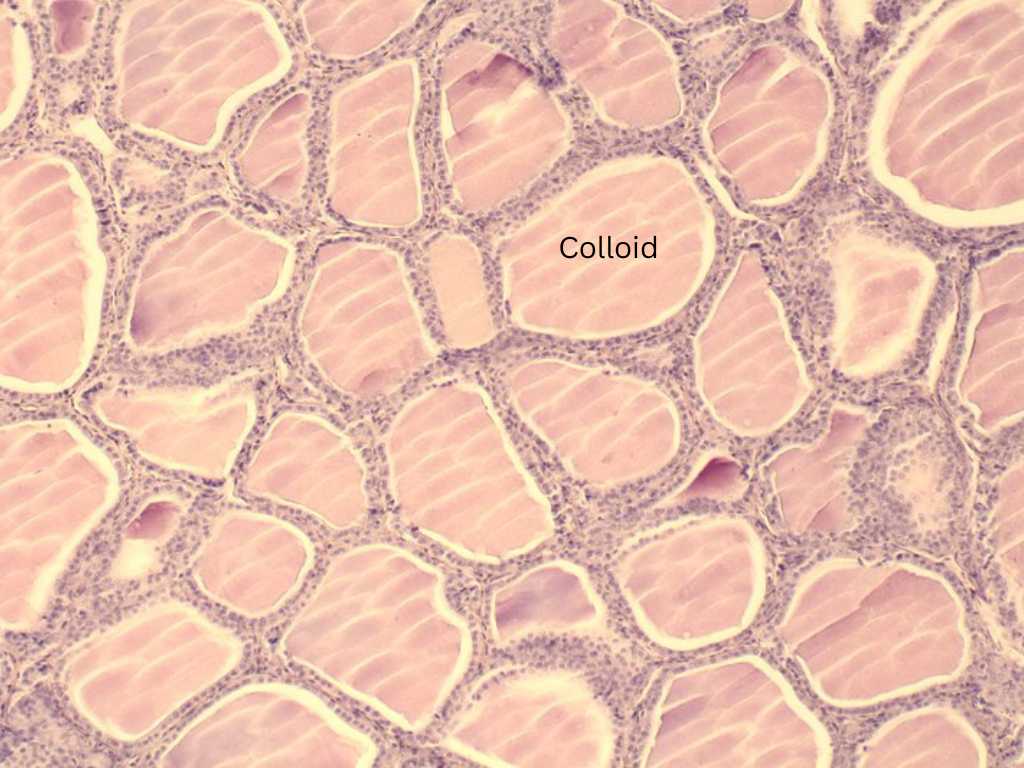
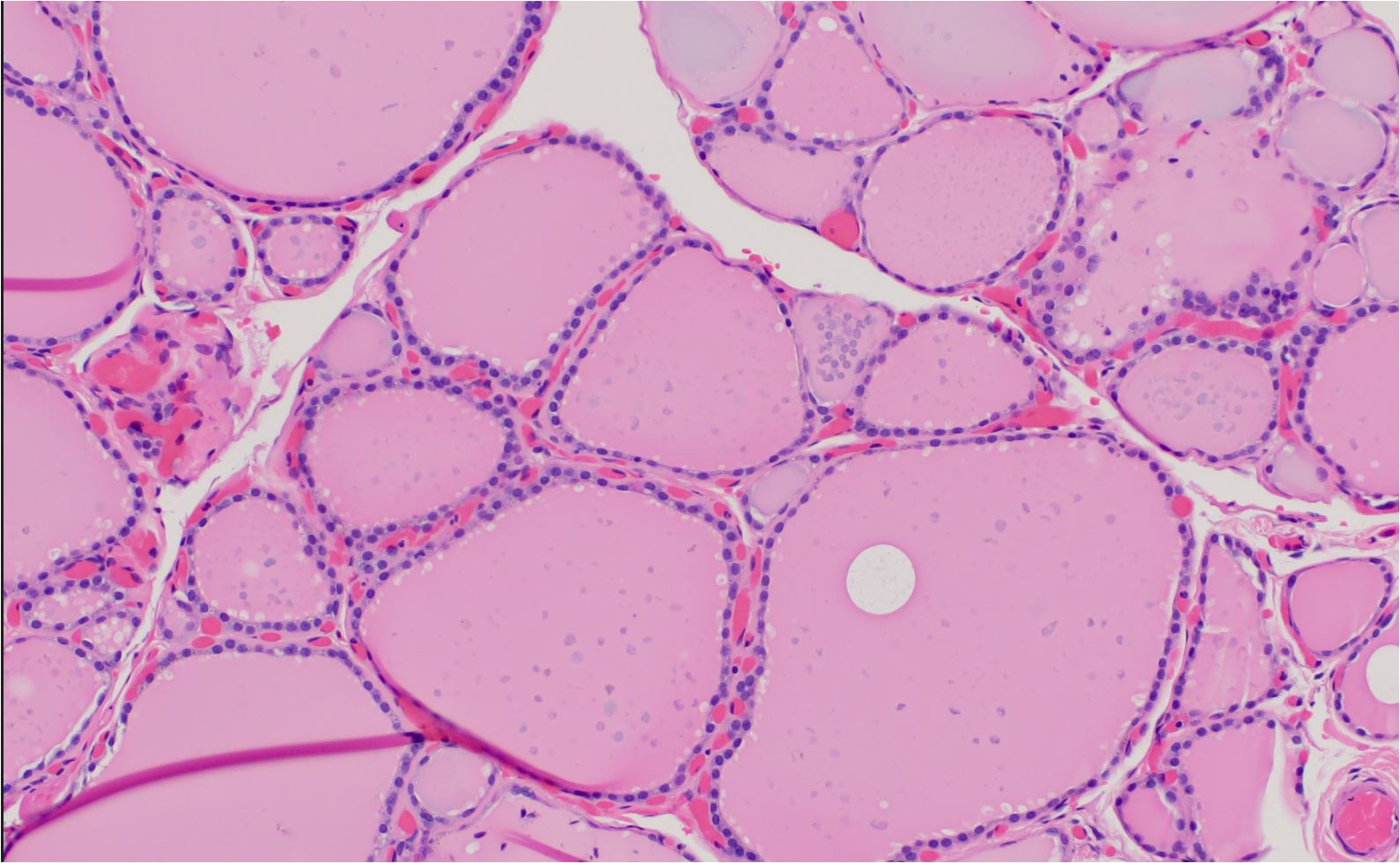
H and E staining of Normal Thyroid Follicular Cells

Normal Thyroid Follicular Cells

Normal Thyroid Follicular Cells (H and E staining at high magnification). Normal thyroid follicular unit lined by a single layer of bland cuboidal follicular cells. Note pink colloid in the lumen
In summary, normal thyroid follicular cells are characterized by their cuboidal or columnar shape, central nucleus, eosinophilic cytoplasm, microvilli on the apical border, and location lining the thyroid follicles. These cells are responsible for the synthesis and secretion of thyroid hormones and play a crucial role in the body's metabolic regulation.
Kindly Let Us Know If This Was helpful? Thank You!


