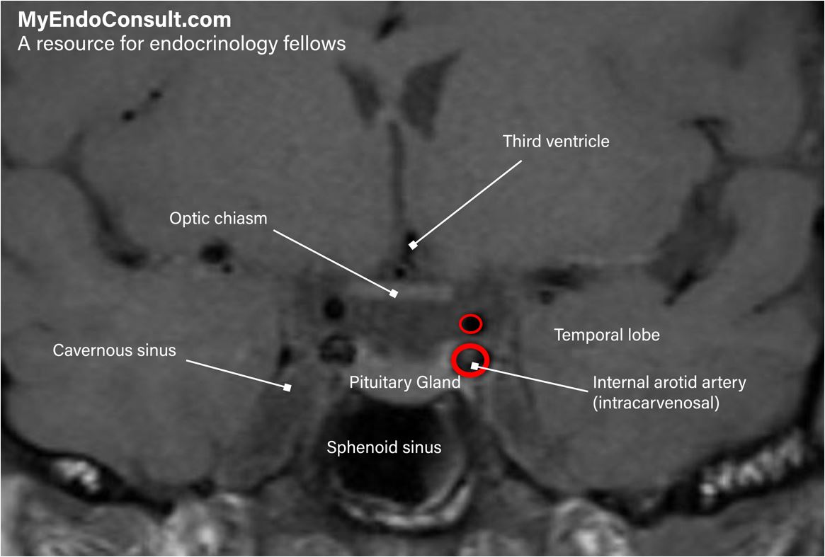
ANTERIOR PITUITARY HORMONES AND FEEDBACK CONTROL
Triple-Level Control
- Hypothalamic Regulation
- Releasing and inhibiting hormones (via the hypophysial portal circulation) act on G-protein–linked receptors in the pituitary, altering gene transcription and hormone secretion.
- Target Gland Feedback
- Circulating hormones from target glands (e.g., cortisol, thyroid hormone) exert negative feedback on their respective pituitary trophic hormones.
- Intrapituitary Regulation
- Autocrine and paracrine cytokines/growth factors locally modulate pituitary cell development and function.
Hypothalamic Releasing/Inhibiting Hormones
- Releasing Hormones: GHRH, CRH, TRH, GnRH.
- Inhibitory Hormones: Somatostatin (inhibits GH) and Dopamine (inhibits prolactin).
Anterior Pituitary Hormones
- Corticotropin (ACTH)
- Growth Hormone (GH)
- Thyrotropin (TSH)
- Follicle-Stimulating Hormone (FSH)
- Luteinizing Hormone (LH)
- Prolactin
- All are secreted pulsatilely into the systemic circulation.
Hypothalamic–Pituitary–Target Gland Feedback Loops
- Negative Feedback: Target gland hormones inhibit further pituitary/hypothalamic secretion (most common).
- Positive Feedback: The target gland hormone or a surrogate may enhance pituitary/hypothalamic release (less common).
- Closed Loop vs. Open Loop:
- Closed Loop: Involves only the trophic hormone and its target gland hormone.
- Open Loop: Includes influence from the nervous system (emotional or external stimuli) that can adjust or override the standard feedback.
Other Factors in Feedback
- Inhibin: A glycoprotein from Sertoli (testes) or granulosa (ovary) cells provides negative feedback on FSH secretion.
- Endogenous Secretory Rhythms:
- Circadian / Diurnal Rhythms: ~24-hour cycle.
- E.g., GH and prolactin peak after sleep onset; cortisol peaks ~2–6 AM, lowest ~11 PM; testosterone highest in the morning.
- Ultradian Rhythms: Occur more frequently than once a day.
- Infradian Rhythms: Periods longer than a day (e.g., menstrual cycle).
- Circadian / Diurnal Rhythms: ~24-hour cycle.
Clinical Considerations
- Pulsatile Secretion: A single GH measurement may not reflect overall GH status; IGF-1 is a more stable index.
- Time-of-Day Reference Ranges: E.g., cortisol reference range differs between morning and afternoon blood draws.
- Loss of Normal Rhythms: E.g., high midnight cortisol supports the diagnosis of Cushing syndrome (disrupted circadian rhythm).
POSTERIOR PITUITARY GLAND
Anatomy and Embryology
- Neural Tissue: Formed by the distal axons of neurons in the supraoptic (SON) and paraventricular (PVN) nuclei of the hypothalamus.
- Hormones Stored: Vasopressin (ADH) and Oxytocin.
- Vasopressin & Oxytocin: Nonapeptides (6–amino acid ring with disulfide bridge + 3–amino acid tail).
- Blood Supply: Inferior hypophysial arteries, draining into the cavernous sinus → internal jugular vein.
Storage and Release
- Storage Capacity: Enough vasopressin to sustain basal release ~30 days and maximal release ~5 days.
- Axonal Transport: ~90% of SON neurons produce vasopressin; the PVN also makes other peptides (somatostatin, CRH, TRH, opioids).
- Secretion: An action potential from SON/PVN travels down the axon, causing Ca²⁺ influx and exocytosis of neurosecretory granules into fenestrated capillaries in the posterior pituitary.
Posterior Pituitary Bright Spot
- MRI Feature: T1-weighted hyperintensity reflecting vasopressin stored in neurosecretory granules.
- Absent in central diabetes insipidus.
- Ectopic Location: In some congenital cases, the bright spot is found along the stalk or at the hypothalamic base; can be associated with a hypoplastic anterior pituitary and variable anterior pituitary dysfunction.