MANIFESTATIONS OF SUPRASELLAR DISEASE

Common Etiologies

  • Tumors / Mass Lesions
    • Craniopharyngioma
    • Dysgerminoma (germinoma)
    • Glioma (e.g., hypothalamic, optic nerve, third ventricle)
    • Meningioma
    • Suprasellar extension of a pituitary tumor
    • Sellar chordoma
    • Hamartoma
    • Gangliocytoma
    • Ependymoma
    • Metastatic neoplasm
  • Cystic / Inflammatory / Granulomatous Lesions
    • Suprasellar arachnoid cyst
    • Lymphocytic hypophysitis
    • Granulomatous diseases (sarcoidosis, tuberculosis, Langerhans cell histiocytosis, Wegener granulomatosis)

Endocrine and Non-Endocrine Sequelae

  1. Visual Pathway Involvement
    • Proximity to the optic chiasm → potential vision loss (bitemporal hemianopsia or other field defects), headaches, and recurrent emesis.
  2. Hypothalamic Dysfunction
    • The hypothalamus controls appetite, sleep–wake cycle, water metabolism, thermoregulation, anterior pituitary function, circadian rhythms, and autonomic inputs.
    • Mass lesions may disrupt one or multiple hypothalamic regions (preoptic, supraoptic, tuberal, mammary) or zones (periventricular, medial, lateral).
  3. Examples of Specific Regional Involvement
    • Posterior Hypothalamus (Mammary Region): Damage → hypersomnolence.
    • Anterior Hypothalamus (Preoptic Region): Hyperactivity, insomnia, altered sleep–wake cycle, dysthermia (acute hyperthermia or chronic hypothermia).
    • Ventromedial Hypothalamus: Lesions → hyperphagia, obesity (destruction of the satiety center).
    • Lateral Hypothalamus: Lesions → hypophagia, weight loss, cachexia (destruction of feeding centers).
  4. Diabetes Insipidus (DI)
    • From destruction of vasopressin-producing magnocellular neurons (supraoptic and paraventricular nuclei) or disruption of the pituitary stalk.
    • Polydipsia/hypodipsia from damage to osmoreceptors in anterior medial/lateral preoptic regions.
  5. Anterior Pituitary Dysfunction
    • Lesions of the tuberal region / floor of the third ventricle → secondary hypothyroidism, secondary adrenal insufficiency, secondary hypogonadism, and GH deficiency.
    • Hyperfunction Syndromes: Certain hypothalamic tumors (hamartomas, gangliocytomas, germ cell tumors) may secrete releasing hormones (e.g., GnRH → precocious puberty; CRH → Cushing syndrome; GHRH → acromegaly).
Pituitary Gland and its Function

CRANIOPHARYNGIOMA

Overview

  • Epidemiology: Most common tumor in the pituitary region in children/adolescents; ~3% of all intracranial tumors (~10% of childhood brain tumors).
  • Pathology: Benign epithelioid tumor from squamous remnants of Rathke pouch. Can be large (>6 cm), often suprasellar, sometimes invading the third ventricle.
  • Location: Usually above the sella turcica, compressing the optic chiasm; can also be intrasellar, eroding the sella floor.

Clinical Presentation

  • Mass Effect
    • Vision loss (chiasm compression)
    • Diabetes insipidus (hypothalamic/stalk invasion)
    • Hypothalamic dysfunction (obesity, hyperphagia, hypersomnolence, temperature regulation disturbances)
    • Anterior pituitary insufficiency (GH deficiency, hypogonadism, adrenal insufficiency, hypothyroidism)
    • Hyperprolactinemia (stalk effect)
    • Signs of increased intracranial pressure (headache, projectile vomiting, papilledema)
    • Hydrocephalus (obstructed CSF flow)
    • Cranial nerve palsies (cavernous sinus involvement)
Anatomical relations of the pituitary gland

Imaging and Histology

  • Skull Radiographs/CT: Irregular suprasellar calcifications.
  • MRI: Multilobulated cystic structure (often cholesterol-rich fluid). May be primarily suprasellar or extend intrasellar.
  • Pathology: Whorls/cords of epithelial cells in a loose stellate network, sometimes with keratohyalin (adamantinomatous variant).

Treatment and Prognosis

  • Options: Observation (in selected cases), transsphenoidal or craniotomy resection, stereotactic radiotherapy, or combinations thereof.
  • Complications: Post-treatment anterior/posterior pituitary hormone deficits are common.
  • Recurrence: ~40% due to tumor adherence to surrounding structures, requiring long-term follow-up.

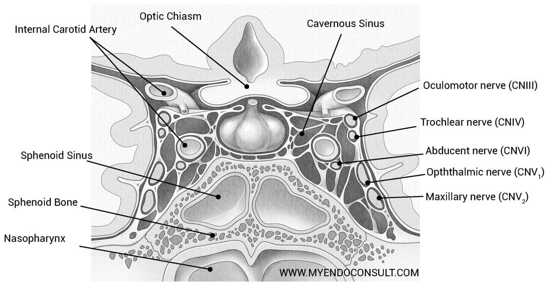
EFFECTS OF PITUITARY TUMORS ON THE VISUAL APPARATUS

Typical Visual Disturbance

  • Bitemporal Hemianopsia
    • Most frequent result of suprasellar extension.
    • Tumor compresses the crossing nasal fibers at the central chiasm.

Variations in Chiasmal Position

  • Prefixed / Postfixed / Lateral Displacements
    • Different chiasm positions can alter the pattern of visual field defects (e.g., homonymous hemianopsia if optic tract is compressed, bilateral central scotomas if posterior chiasm is affected).
  • Other Visual Defects
    • Unilateral central scotoma, amblyopia in one eye, or inferior quadrantanopia if specific chiasm regions or optic pathways are compressed.

Recovery and Limitations

  • If the pressure is relieved surgically or medically, vision may improve depending on the degree/duration of tract compression.
  • A tough diaphragma sellae or higher position of the chiasm may delay onset of visual symptoms but may allow lateral or inferior tumor extension.

NONTUMOROUS LESIONS OF THE PITUITARY GLAND AND STALK

Etiologies

  1. Lymphocytic Hypophysitis
    • Autoimmune; often in late pregnancy or postpartum.
    • Causes pituitary enlargement, headaches, hormone deficiencies (often ACTH deficiency).
    • MRI: Homogeneous, enhancing sellar mass that may involve the stalk.
    • Variable recovery; some have permanent deficits, others regain partial function.
  2. Granulomatous Disorders
    • Sarcoidosis, Tuberculosis, Langerhans Cell Histiocytosis, Wegener Granulomatosis
    • Can involve hypothalamus, pituitary stalk, gland → hypopituitarism (including DI).
  3. Head Trauma
    • Skull base fracture, stalk section, traumatic vasospasm → pituitary infarction.
    • Leads to anterior/posterior pituitary failure of varying degrees.
  4. Iron Overload
    • Hemochromatosis, hemosiderosis → iron deposition in pituitary (siderosis), often selectively impairing gonadotropin secretion.
  5. Empty Sella Syndrome
    • Secondary: Post-surgical, post-radiation, or post-infarction enlargement of the sella with no substantial gland filling it.
    • Primary: Defect in diaphragm sellae allows CSF to enter/enlarge the sella (may accompany benign intracranial hypertension).
    • Often pituitary function remains intact, with the gland compressed against the sellar floor.
  6. Genetic/Developmental Anomalies
    • Transcription Factor Mutations (HESX1, LHX3, LHX4, PROP1, POU1F1, TBX19) → combined pituitary hormone deficiencies.
    • Midline Anomalies: Cleft palate, encephalocele, optic nerve hypoplasia; can cause pituitary aplasia/hypoplasia or ectopic gland.

Join the
MyEndoConsult Community

We are grateful to the contribution of authors just like you

The MyEndoconsult Team. A group of physicians dedicated to endocrinology and internal medicine education. Learn more about our team

Current Progress
Current Progress
Current Progress
Current Progress
>