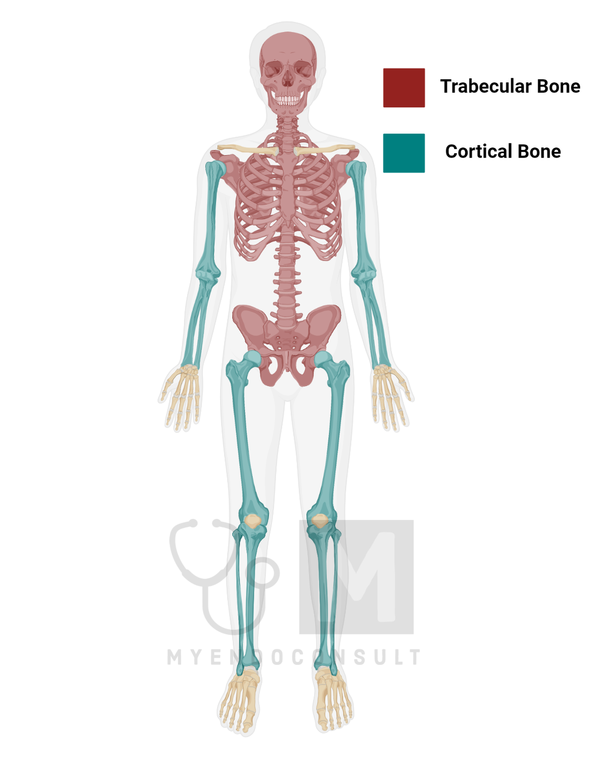
Distinct types of bone tissue can be recognized visually — dense, compact bone tissue (cortical bone) and porous, spongy bone tissue (trabecular bone).
The dense version, known for its low proportion of soft tissue, under 10%, contrasts markedly with the spongy component, which is structured with rod-like or plate-like structures known as trabeculae, amid significant bone marrow that amounts to over 75% of the spongy bone’s volume.
The dense bone tissue is the outermost layer of all bones and is chiefly concentrated in long bones (limbs). Conversely, the porous version primarily resides in the central skeleton, taking up space between the denser bone of smaller, flat, and short bones like shoulder blades, vertebrae, and pelvic bones. It can also be found, although sparingly, at the ends near the joints of limb bones.
Even though the dense bone tissue comprises 80% of the total bone mass, providing the majority of the mechanical function, the spongy bone, representing only 20% of the bone mass, displays impressive metabolic activity. In fact, it is metabolically four times more active per volume unit compared to the dense bone.
Surfaces of Bone
Bones at the microscopic scale exhibit a couple of protective layers. The external periosteal layer is a connective tissue that encapsulates rigid and soft tissues while isolating the bone from surrounding organs. The internal endosteal layer encompasses all soft tissue inside the bone, excluding osteocytes. This layer demarcates the boundary between bone marrow, soft tissue, and bone tissue.
Within this endosteal layer, we can identify three unique yet interconnected surfaces: the intracortical surface, including certain biological canals; the endocortical surface; and the trabecular surface. The activity of bone modeling and remodeling happens on these surfaces. Nevertheless, the intensity of these activities can differ among surfaces and can be variably impacted by physiological or pathological factors and treatment methods.
What is the Bone Structural Unit
Concerning the Bone Structural Unit, within the boundaries set by the periosteal and endosteal layers, bone is organized into several structural components, referred to as bone structural units (BSUs). The layout and cohesion of these minute bone units contribute to the bone’s overall strength.
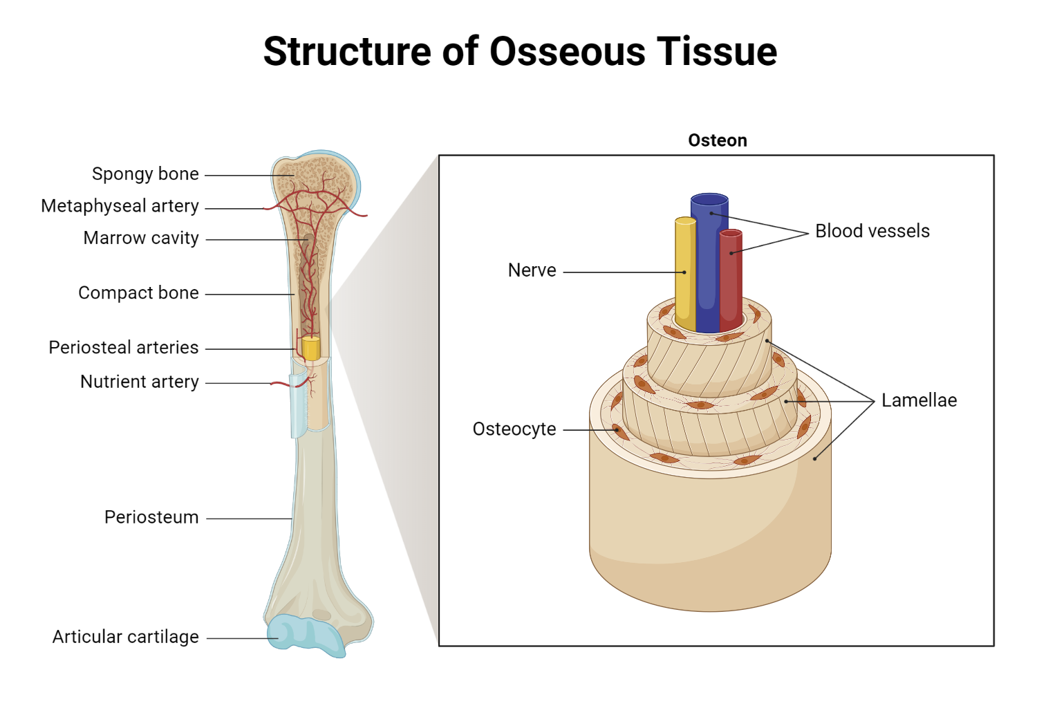
In the case of cortical bone, the osteon or haversian system represents the BSUs. Each osteon is a cylindrical structure aligned with the long axis of the cortical bone. The center of the osteon is taken up by a haversian canal, housing blood vessels, nerves, and connective tissue. These canals from neighboring osteons are interconnected laterally by Volkmann’s canals, giving rise to an intracortical network. This network is also linked with the periosteum and bone marrow. The central haversian canal is circled by around 20 to 30 bone lamellae, forming the osteon wall. The osteons are packed closely together, separated only by the remains of partially resorbed osteons, also known as interstitial lamellae.
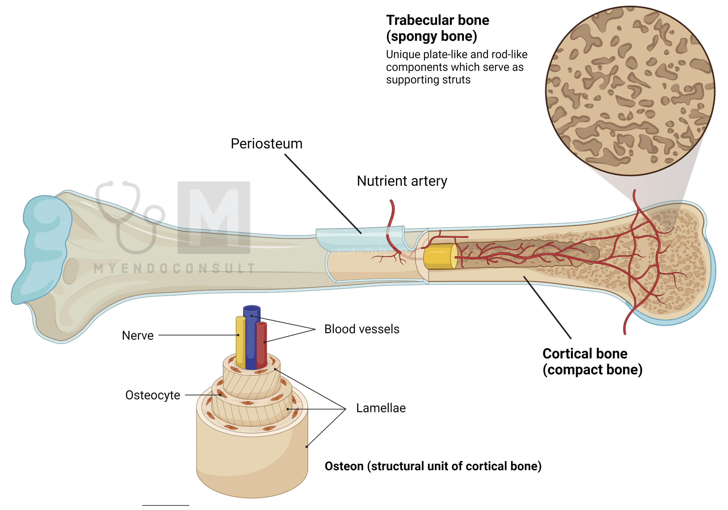
In spongy or cancellous bone, the bone structural units (BSUs) are flat and could be likened to osteons longitudinally sliced open and unfolded. They form a crescent-shaped structure, roughly running parallel to the central axis of the trabeculae, a small rod or plate-like piece of bone tissue. The trabecular surface, which corresponds to the open haversian canal, conforms to the shape of the trabecule and interfaces with the bone marrow. The BSUs within the trabecules are separated from one another by interstitial bone. Much like in cortical bone, this interstitial bone constitutes the remnants of older partially absorbed units.
Each Bone Structural Unit (BSU) comprises a specialized connective tissue, the osseous tissue, which is made up of a mineralized protein matrix. On a microscopic scale, the arrangement of collagen bundles distinguishes two different types of bone.
In a typical mature skeleton, the bone is of the lamellar type. In this type of bone, the direction of collagen fibers alternates consistently from one layer to another. Each layer is approximately 3 micrometers thick, and all collagen fibers within the same layer are deposited in the same direction. The lamellar pattern of collagen deposition is quite orderly. The layers that are circular alternate with the longitudinal ones. This changing direction of collagen fibers from layer to layer accounts for bone’s birefringence, or light-refracting properties, when observed under polarized light microscopy.
In contrast to the regularity of lamellar bone, woven bone consists of loosely and randomly organized collagen bundles. This form of bone is the result of the irregular, unpolarized excretion of protocollagen by osteoblasts, or bone-forming cells. The matrix displays a disorganized, crisscross pattern and lacks the characteristic birefringence of lamellar bone under polarized light.
Woven bone is found in the embryonic skeleton and in both cortical and cancellous bones during periods of rapid growth. After bone growth is complete, woven bone in the normal skeleton is replaced by lamellar bone. However, woven bone is also noted in specific pathological conditions, such as Paget’s disease of the bone, bone fracture healing, osteogenesis imperfecta, and in both primary and secondary hyperparathyroidism. In adults, the presence of woven bone signifies rapid, unregulated bone formation and high bone turnover, which could be due to local or systemic factors.
Comparison of Cortical and Trabecular Bone
| Feature | Cortical Bone | Trabecular Bone |
| Location | Found on the outer surface of the bone, making up the hard outer layer | Found inside the ends of long bones, in the pelvis, ribs, skull, and in the vertebrae in the spinal column |
| Structure | Solid and dense; compact | Spongy, less dense, and honeycomb-like |
| Percent of skeleton | Makes up about 80% of the skeletal mass | Makes up about 20% of the skeletal mass |
| Function | Provides support and is resistant to bending and torsion | Provides strength while also being lighter, reduces the load on the bones by transferring loads to other parts of the bone |
| Blood supply | Poorly vascularized | Highly vascularized |
| Remodeling rate | Slower remodeling rate due to denser structure | Faster remodeling rate due to its spongy nature |
| Cells found | Osteons are densely packed together | Contains trabeculae that provide room for bone marrow and blood vessels |