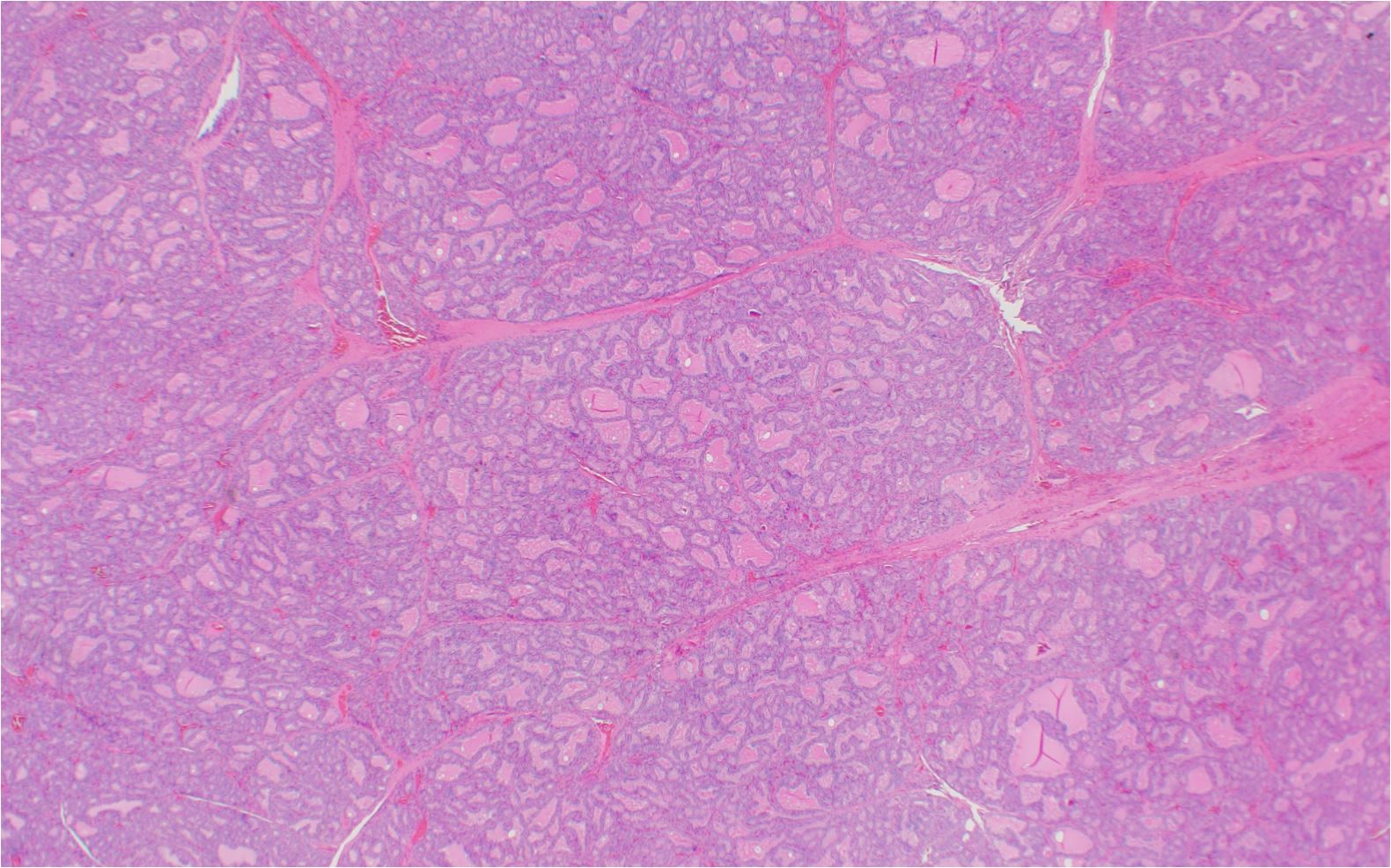
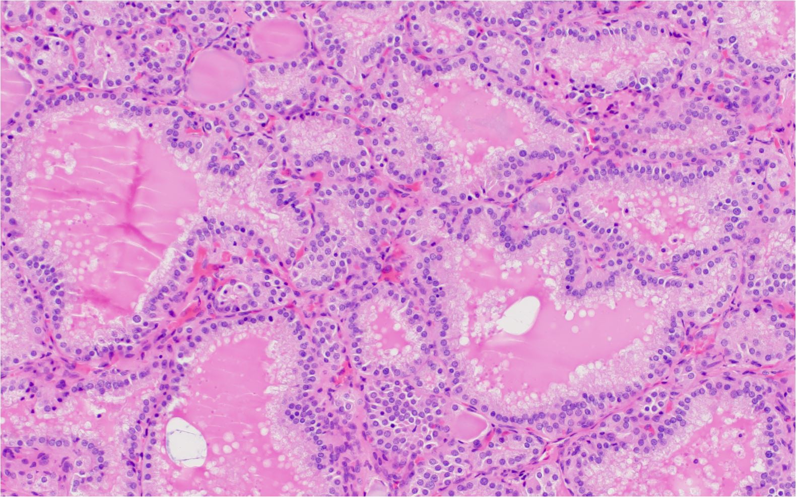
Graves' disease is an autoimmune condition that affects the thyroid gland, leading to hyperthyroidism, which is characterized by an overproduction of thyroid hormones. It is the most common cause of hyperthyroidism and occurs more frequently in women than in men. In this review, we will provide a detailed description of the histological features of Graves' disease.

Follicular hyperplasia and lymphoplasmacytic infiltrate clinically associated with Graves disease. Lymphoid follicle on right side Papillary infoldings (arrows)
Histological features of Graves Disease
Follicular architecture: The thyroid follicles in Graves' disease are often irregularly shaped and variable in size. Furthermore, they may appear small, compressed, and elongated, with a reduction in colloid content.
Indeed, the colloid may appear patchy or scalloped, and in some cases, it can be nearly absent.
Follicular cells: The thyroid follicular cells in Graves' disease are usually hyperplastic and may appear taller (columnar) than the typical cuboidal shape of normal follicular cells.
It is worthy to note that this change in morphology reflects the increased activity of these cells in producing thyroid hormones.
Stromal changes: The stroma in Graves' disease may exhibit infiltration by inflammatory cells, including lymphocytes, plasma cells, and macrophages.
The presence of lymphoid aggregates, which are clusters of lymphocytes, may be observed, and germinal centers can occasionally form.
This pattern of lymphocytic infiltration is a hallmark of the autoimmune nature of the disease.
Vascularity: Increased vascularity is a characteristic attribute of Graves' disease. It is worthy to note that the blood vessels within the thyroid gland may be more prominent, dilated, and congested due to the increased metabolic demand and thyroid hormone production.
Capsule: In some cases, the thyroid capsule in Graves' disease may be thickened and fibrotic. This thickening can be attributed to inflammation, as well as fibrosis resulting from long-standing disease.
Ophthalmopathy: Graves' disease may be associated with exophthalmos, or bulging of the eyes, due to the infiltration of the orbital tissues by inflammatory cells, glycosaminoglycans, and fluid.
This condition, known as Graves' ophthalmopathy or thyroid eye disease, can cause various ocular symptoms, including eye pain, dryness, redness, and vision changes.
H and E features of Graves Disease

Diffuse follicular hyperplasia with infoldings of the follicular epithelium. Active tall follicular cells. Decreased colloid. Peripheral scalloping of colloid (arrow). Lymphoid infiltrate (lower right corner)

Diffuse folllicular hyperplasia without inflammatory infiltrate clinically associated with graves disease

Folllicular hyperplasia without inflammatory infiltrate clinically associated with graves disease. Peripheral scalloping of colloid
In summary, the histological features of Graves' disease include diffuse enlargement of the thyroid gland, irregular and variable-sized thyroid follicles with reduced colloid content, hyperplastic and columnar follicular cells, infiltration by inflammatory cells, increased vascularity, and occasional thickening of the thyroid capsule.
These histologic attributes help to distinguish Graves' disease from other causes of hyperthyroidism.
Kindly Let Us Know If This Was helpful? Thank You!


