Learning Objectives
- Understand the basic anatomy of the thyroid gland and its structural relations in the central compartment of the neck
- Understand the appearance of a normal thyroid gland on ultrasonography.
- Appreciate important ultrasound features (pattern recognition) of anatomic relations of the thyroid gland.
Anatomy of the thyroid gland and relevant cervical structures
Thyroid, which in Greek means “shield-shaped,” is a term credited to Bartholomeus Eustachius of Rome. He aptly described the thyroid gland as “a single glandulamthyroideam” with two lobes connected via ridge (or isthmus). The normal thyroid gland hugs the trachea anterolaterally and is bound laterally by the carotid sheath (containing the common carotid artery, internal jugular vein and vagus nerve) and sternomastoid muscles[1].
The strap muscles (sternohyoid, sternothyroid, and omohyoid) cover most of the anterior aspect of the thyroid gland.
The gland usually measures anywhere from 15 to 25grams in weight. The lateral lobes are about 4cm in length and 2cm in width.
The isthmus (which connects both lateral lobes) crosses the trachea in front of the first two tracheal rings.
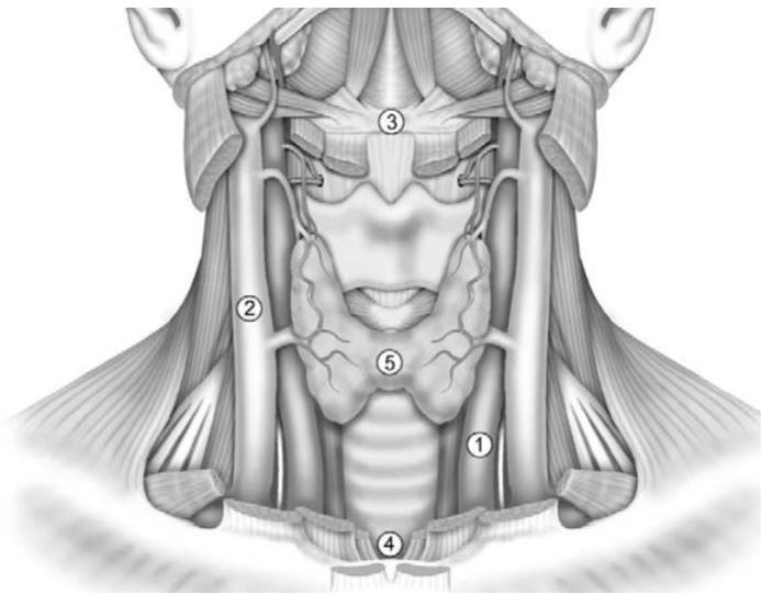
Figure 1.0 Anatomy of the central compartment of the neck, depicting important relations of the thyroid gland. The carotid artery(2) and internal jugular vein (1) lying laterally, hyoid bone (3) superiorly, and suprasternal notch (4) inferiorly. Based on Gavilian et al [2]
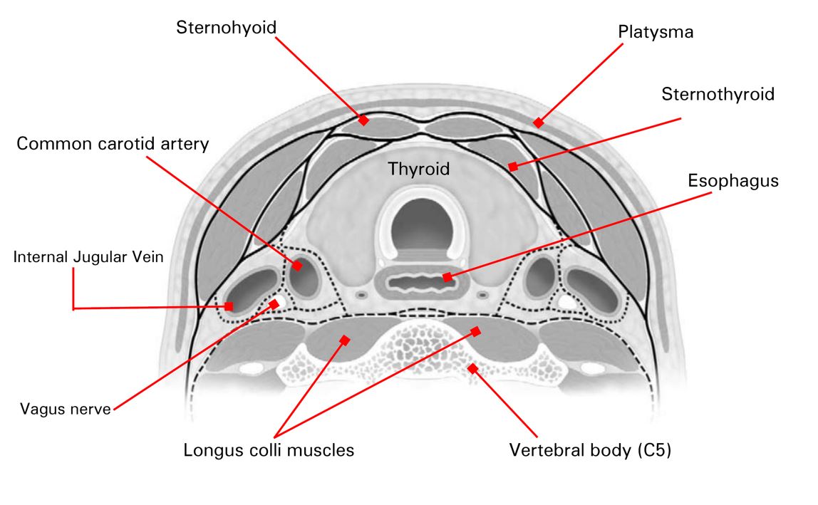
Figure 2.0. A schematic representation of the transverse cross-section of the neck at the level of the mid-thyroid gland. Deep cervical fascia encasing neck muscles is shown as a solid black line. The carotid sheath is shown as a dotted line, and the prevertebral fascia by a dashed line. Anatomic relations of the thyroid are shown. Adapted from Gavilian et al [2]
Findings on thyroid ultrasonography
Due to its superficial location, the thyroid is easily discernible on ultrasonography.
Traditional piezoelectric ultrasound transducers
Ultrasound transducers are composed of an array of piezoelectric crystals which play the dual role of transmitting and receiving ultrasound energy. These crystals vibrate when exposed to electric current, generating ultrasound energy. Conversely, reflected ultrasound energy heading back to the transducer from both superficial and deep tissues strikes these crystals and initiates electrical energy generation.
Sound waves are transmitted as mechanical energy and require a medium to propagate. Sound waves are therefore unable to traverse a vacuum—the density and stiffness of the medium impact the speed of transmission of sound waves.
Acoustic impedance is defined as the inverse of the capacity of a medium to transmit sound. Indeed, when sound waves change from one medium to another, a change in acoustic impedance leads to a partial reflection of sound waves.
Subsequent capturing of these reflected waves followed by image reconstruction and enhancement results in the visualization of sharp images of the thyroid and its anatomic relations.
Modern chip-based ultrasound devices
Chip-based ultrasound devices have revolutionized point-of-care ultrasonography. The chips have multiple “drums” which effectively behave like piezoelectric crystals by wobbling (or vibrating) to produce ultrasound waves under direct stimulation by electrical energy. In addition, these chips receive reflected sound energy and convert it into electric energy that can then be rendered into an image on a smartphone or tablet. These chip-based transducers have a much wider bandwidth than traditional piezoelectric transducers, which allows a single probe to be utilized at various sites, including the thyroid, cardiac, bladder, kidney, liver etc.
Video clip depicting the left thyroid lobe captured using the butterfly IQ® chip-based transducer. Video courtesy of Dr. A. Quarde
The use of higher frequency probes (as high as 16MHz) provides more excellent resolution of the thyroid gland in its superficial position but limits the depth of penetration (typically less than 5cm).
Color-flow doppler provides visualization of both the speed and direction of blood flow within the soft tissue of the thyroid gland[3].
The appearance of the normal thyroid gland on ultrasound
The normal thyroid gland has a ground glass appearance and is brighter (hyperechoic) than the strap muscles (sternohyoid and sternothyroid).
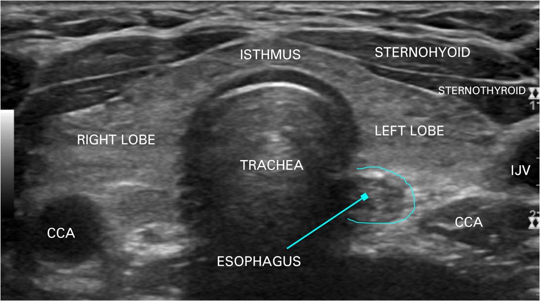
Figure 3.0 Transverse view of a normal thyroid gland showing important anatomic relations. CCA common carotid artery, IJV internal jugular vein. Image courtesy of Dr. A. Quarde
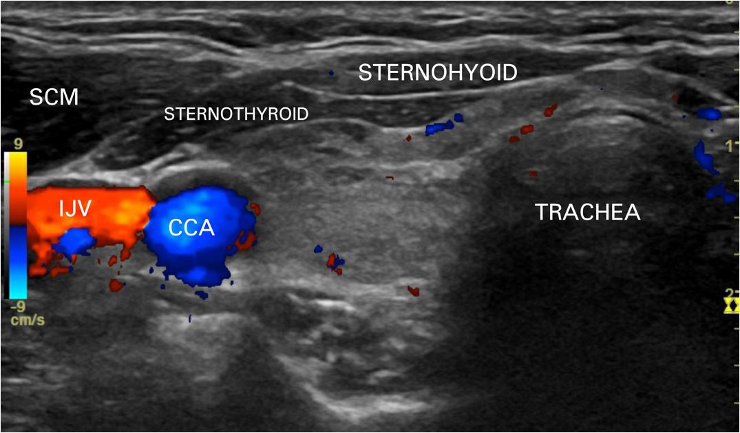
Figure 4.0. Transverse view of the right thyroid lobe. CCA common carotid artery, IJV internal jugular vein, SCM sternocleidomastoid/sternomastoid muscle. Image courtesy of Dr. A. Quarde
References
- Chaudhary V, Bano S (2013) Thyroid ultrasound. Indian J Endocrinol Metab 17:219–227
- Gavilán J, Herranz J, Martín L (2004) Functional neck dissection: The Latin approach. Operative Techniques in Otolaryngology-Head and Neck Surgery 15:168–175
- Fukunari N (2002) Thyroid ultrasonography B-mode and color-Doppler. Biomed Pharmacother 56 Suppl 1:55s–59s