Salt and pepper skull is a radiographic finding that originates from famous mid-17th-century French cuisine, highlighting the importance of white salt and black pepper as essential condiments at the dinner table [1].
Literally, white salt refers to osteosclerotic areas, while black pepper refers to lytic areas of bone noted on a lateral skull radiograph (x-ray).
Radiographic description of salt and pepper skull
This is classically described as discrete areas of both osteolytic (spotty deossification) and osteosclerotic changes typically noted on the lateral skull radiograph of patients with elevated levels of serum PTH (hyperparathyroidism of various causes). It is the ultimate mimic of skull metastases and may lead to a presumptive diagnosis of cancer in the unsuspecting clinician with minimal experience in diagnosing this condition.
This radiographic finding is found in patients with osteitis fibrosa cystica (OFC), a protean skeletal manifestation of hyperparathyroidism of any etiology. Salt and pepper appearance of the skull (sometimes called, pepper pot skull), bone cysts, brown tumors of long bones, and subperiosteal resorption of the distal phalanges are usual radiological features of osteitis fibrosa cystica. These findings are now seldom seen in practice due to an improvement in diagnosis and management of hyperparathyroidism.
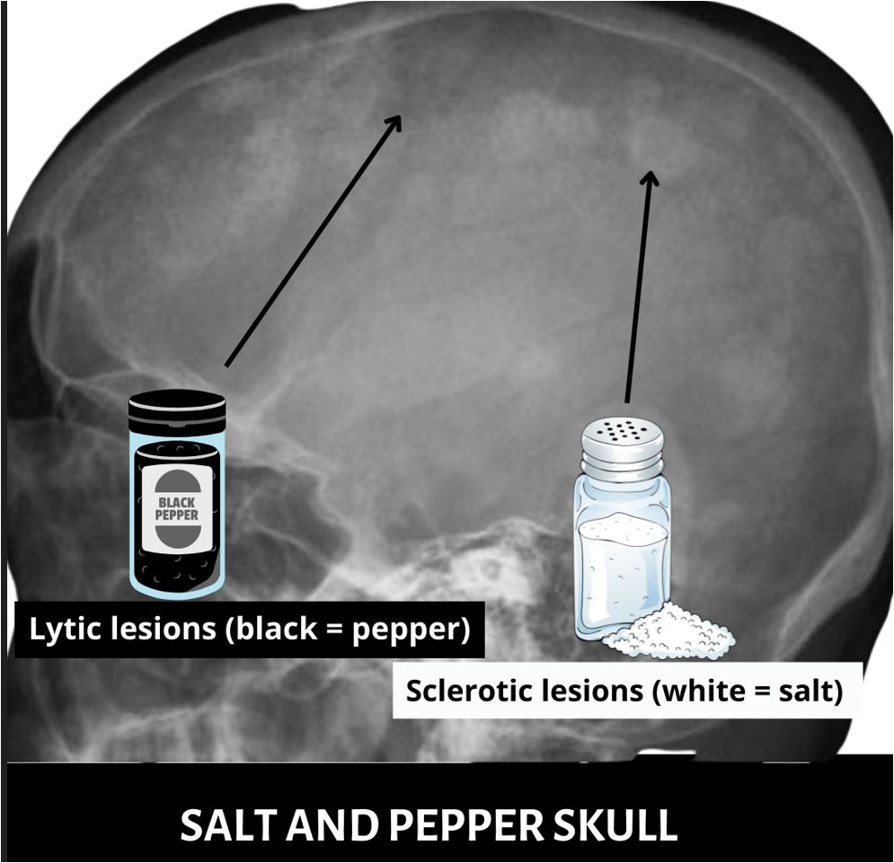 salt and pepper skull (lateral skull radiograph)
What causes salt and pepper skull?
Various causes of primary, secondary, or tertiary hyperparathyroidism can result in a salt and pepper appearance of the skull. Primary hyperparathyroidism occurs as a result of parathyroid adenoma in more than 85% of patients.
Secondary hyperparathyroidism refers to diffuse 4 parathyroid gland hyperplasia in the setting of end-organ resistance to PTH action. This is usually seen in patients with chronic kidney disease. The progressive loss of renal mass causes a steady decline in glomerular filtration rate. Consequently, there is reduced phosphate excretion and 1-alpha hydroxylase activity. These metabolic perturbations drive increased parathyroid gland proliferation and parathyroid hormone hypersecretion.
Patients with secondary hyperparathyroidism may experience an improvement in serum PTH and calcium levels if the cause of end-organ resistance to PTH action is corrected.
In patients with a long-standing history of secondary hyperparathyroidism, either one or more of the hyperplastic parathyroid may achieve autonomy. This means either one or more hyperplastic parathyroid glands will independently release parathyroid hormone irrespective of serum calcium concentration, essentially behaving like a parathyroid adenoma.
Additional causes of tertiary hyperparathyroidism include chronic vitamin d deficiency, hypophosphatemic osteomalacia, CKD and phosphate supplementation with concomitant calcitriol.
The underlying pathophysiology of salt and pepper skull
Although the pathophysiology of salt and pepper skull is unknown few possible mechanisms have been proposed. It is worthy to note that remodeling-based bone formation occurs after a period of prolonged osteoclastic activity. Hyperparathyroidism promotes parathyroid hormone-mediated bone resorption (dark in appearance, represents pepper).
After a prolonged period of bone resorption, there is a change in the tide, with osteoblastic activity disproportionately higher than osteoclastic activity[2]. This again is mediated by parathyroid hormone action. Subsequently, osteoblast-mediated bone formation leads to osteosclerosis. This appears radiologically as areas of sclerosis (whitish in appearance, represents salt).
Complications
The complications of this condition tend to be an increase in the risk for fractures due to decreased bone mass.
Treatment
treatment involves correcting the underlying cause. Indeed, there have been reports of complete resolution of the salt-and-pepper appearance of the skull after successful treatment of hyperparathyroidism, irrespective of the etiology [3].
What is a raindrop skull?
A raindrop skull sign of calvarial multiple myeloma is characterized by multiple, symmetrical lytic lesions spread around the skull. This term represents a visual pattern of raindrops hitting and splattering on a surface leaving a random pattern of dark spots.
References
1. Peterson TS. HOW SALT & PEPPER TOOK OVER THE WORLD — A FRENCH TALE. Washington Post. https://www.washingtonpost.com/archive/lifestyle/food/1995/12/27/how-salt-pepper-took-over-the-world-a-french-tale/a2a8d70c-5ee6-45a5-8e0f-ec9514c74af1/. Published December 27, 1995. Accessed June 20, 2022.
2. Shetty S, Kapoor N, Naik D, Paul TV. Focal osteosclerosis of the skull in primary hyperparathyroidism. BMJ Case Rep. 2014;2014:bcr2014204236. doi:10.1136/bcr-2014-204236
3. Kaur G, Singh P, Mittal N, Singla MK. Resolution of “salt and pepper” appearance of the skull with vitamin D therapy. Indian J Endocrinol Metab. 2013;17(Suppl1):S194-S197. doi:10.4103/2230-8210.119569