Papillary thyroid carcinoma (PTC) is the most common type of thyroid cancer, accounting for approximately 80% of all thyroid malignancies. PTC typically has an indolent course and a favorable prognosis, with a high overall survival rate. In this review, we will provide a detailed description of the histological features of papillary thyroid carcinoma, including characteristic histologic features such as "Orphan Annie eye" nuclei and psammoma bodies.
Histological Features of Papillary Thyroid Carcinoma

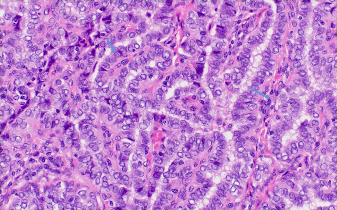
Papillary thyroid carcinoma, classic nuclear features. Marked nuclear enlargement (compare to normal thyroid follicular epithelium). Nuclear crowding Grooves (short arrow). Nuclear condensation to the periphery of the nucleus (Orphan Annie Eye). Papillary architecture characterized by fibrovascular core (long arrow).
- Papillary architecture: The hallmark feature of PTC is the presence of papillary structures, which are finger-like projections of tumor cells supported by a central fibrovascular core. These papillae can vary in size and shape and may be branched or complex.
- Tumor cells: The tumor cells in PTC are usually cuboidal or columnar, with a moderate amount of eosinophilic or clear cytoplasm. Also, the cells typically form a monolayer along the papillary structures and can be arranged in various patterns, such as follicular, solid, or cystic.
- "Orphan Annie eye" nuclei: The presence of ground-glass, optically clear nuclei, often referred to as "Orphan Annie eye" nuclei is a unique distinguishing feature of papillary thyroid carcinoma. These nuclei are large, oval or round, with a pale, glassy appearance due to the margination of chromatin to the nuclear periphery and is highly suggestive of PTC as it is not typically seen in other thyroid neoplasms.
- Nuclear grooves: It is worthy to note that PTC is the presence of nuclear grooves or folds, which are linear invaginations of the nuclear membrane. Actually, these grooves can be seen in a significant proportion of tumor cells and are an important diagnostic clue for PTC.
- Intranuclear cytoplasmic inclusions: Intranuclear cytoplasmic inclusions are occasionally observed in PTC. These are small, eosinophilic cytoplasmic projections that extend into the nucleus and can help confirm the diagnosis of PTC.
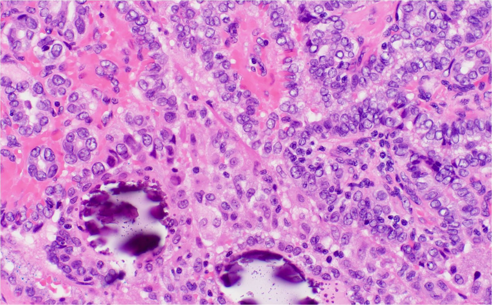
- Psammoma bodies: Psammoma bodies are concentrically laminated, calcified structures often found in PTC. It has been postulated that they arise from the calcification of degenerating tumor cells or from the mineralization of the basement membrane. While not specific to PTC, the presence of psammoma bodies can support the diagnosis when observed in conjunction with other characteristic features.
- Lymphatic invasion and metastasis: PTC frequently exhibits lymphatic invasion and is known for its propensity to metastasize to regional lymph nodes, in contrast to follicular carcinomas, which tend to spread hematogenously. Lymph node metastasis may be present at the time of diagnosis or develop during the course of the disease.
- Capsular invasion: PTC may invade the thyroid capsule, which can be an indicator of more aggressive behavior and a higher risk of recurrence. However, capsular invasion alone is not sufficient to confirm malignancy, as it can also be seen in some benign thyroid lesions.
- Multifocality: PTC can be multifocal, with multiple tumor foci within the same thyroid gland. This finding is associated with a higher risk of recurrence and lymph node metastasis.
H and E Features of Papillary Thyroid Carcinoma

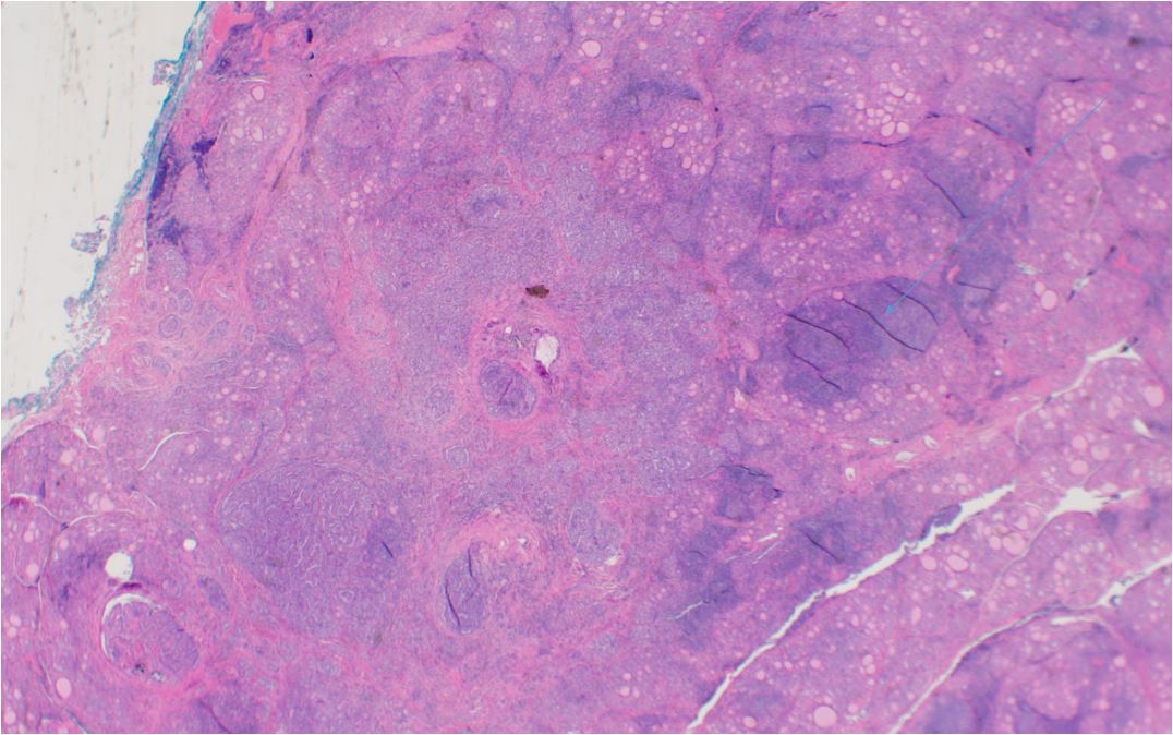
Papillary thyroid carcinoma, conventional-type, occurring in a background of Hashimoto’s thyroiditis. Note sclerosis in area of tumor and lymphoid infiltrated in surrounding thyroid (large arrow)

Papillary thyroid Carcinoma with psammatous calcifications (crushed)
In summary, the histological features of papillary thyroid carcinoma include papillary structures lined by cuboidal or columnar tumor cells, characteristic "Orphan Annie eye" nuclei, nuclear grooves, intranuclear cytoplasmic inclusions, psammoma bodies, lymphatic invasion, and the potential for capsular invasion and multifocality. The presence of these histologic attributes helps to distinguish PTC from other thyroid neoplasms and contribute to its diagnosis and prognostic assessment.
Kindly Let Us Know If This Was helpful? Thank You!


