A thyroid examination is a simple bedside evaluation that will help diagnose thyroid disorders, including hypothyroidism, hyperthyroidism, or both. The thyroid gland is located in your neck below the thyroid cartilage (Adam’s apple). It produces two important hormones: 3,5,3′,5′-tetraiodothyronine (T4) and 3,5,3′-Triiodo-l-thyronine (T3).
There are several different types of thyroid disease, including Hashimoto’s disease (which causes an underactive thyroid gland), Graves’ disease (autoimmune mediated inflammation of the thyroid gland which results in productive hyperthyroidism), or toxic nodular goiter (with benign nodules producing excess thyroid hormone)
Inspection of the thyroid gland
- A visible anterior neck mass, which is mobile on deglutition, implies an enlarged gland. Offer the patient a glass of water to drink. The normal thyroid gland cannot be visualized on inspection.
- Positive Pemberton’s sign is suggestive of significant retrosternal extension of a goiter.
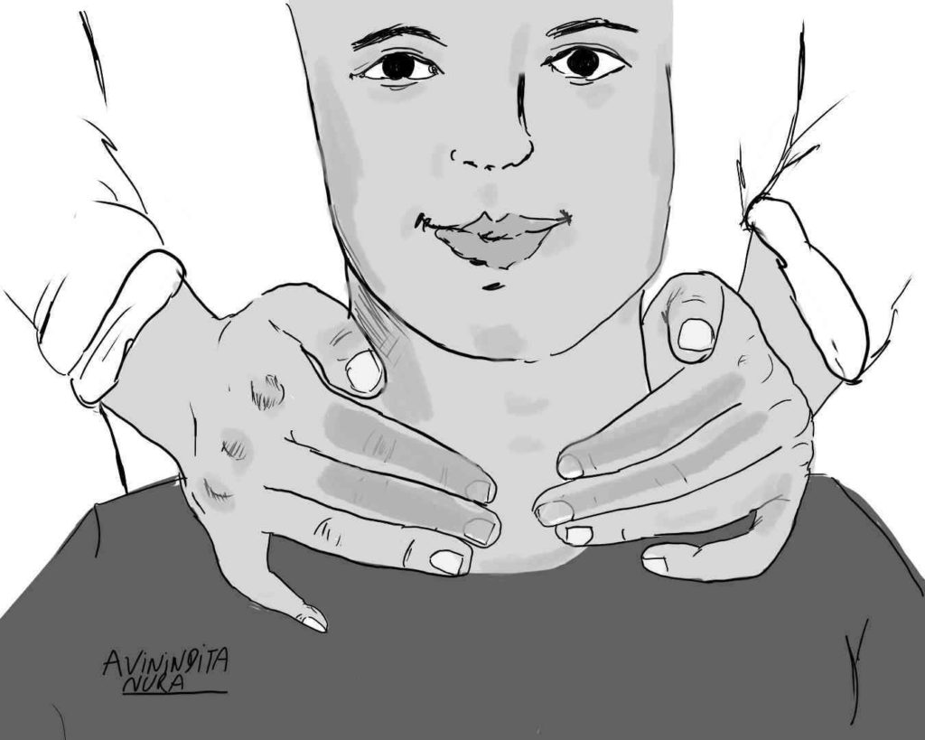
Palpation
- Optimal positioning of the neck will facilitate palpation of the gland. The patient’s neck should be in a relaxed position, which prevents extensive nuchal extension or flexion. The sternocleidomastoid muscle should not be under tension, and there should be adequate room between the chin and the sternum to allow proper positioning of the hands.
- Sequentially palpate each thyroid lobe and isthmus. Occasionally there might be an accessory extension of the isthmus – the pyramidal lobe. Check for the consistency of the gland. Avoid deep palpation if the gland is tender, as might occur in De Quervain’s thyroiditis. The normal thyroid has a mild firm consistency. The overlying skin is usually freely mobile over it. If the gland is attached to overlying skin, it might be suggestive of either a malignancy or Riedel’s thyroiditis.
- Attempt to feel for any discrete thyroid nodule. These are best appreciated using the pulp of your examining fingers (2nd, 3rd, and 4th digits)
- Ask the patient to swallow during palpation and check for any retrosternal extension of the thyroid gland.
- Grade thyromegaly with the WHO classification system
- Palpate all regional lymph node groups (I to VI)
Percussion
- Percussion of the sternal manubrium may be required in patients with a suspected retrosternal goiter. The percussion note will be dull. This step is seldom warranted, especially if there is no palpable thyroid tissue extending below the proximal portion of the manubrium (thoracic aperture).
Auscultation
- In patients with hyperdynamic circulation, as might occur in overt Graves disease, a bruit may be appreciated over the superior poles of the thyroid in the vicinity of the superior thyroid arteries.
Kindly Let Us Know If This Was helpful? Thank You!